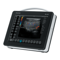
DRAMIŃSKI BLUE – user manual
4
9.1.4. Vi–Probe.................................................................................... 30
10. Description of the ultrasound scanner functions ...................................................... 31
10.1. Optimizing the image parameters ...............................................................31
10.1.1. Gain control ...............................................................................31
10.1.2. Time Gain Control (TGC) ................................................................... 31
10.1.3. Frequency ................................................................................. 31
10.1.4. Depth .....................................................................................31
10.1.5. Focus......................................................................................32
10.1.6. Zoom .....................................................................................32
10.1.7. Dynamic range ............................................................................32
10.1.8. Gamma.................................................................................... 32
10.1.9. D–Curve................................................................................... 33
10.2. Freeze ..........................................................................................33
10.3. Cine loop – continuous and frame-by-frame replay ..............................................33
10.4. Measurements..................................................................................33
10.4.1. Length ....................................................................................34
10.4.2. Grid .......................................................................................34
10.4.3. Stenosis ...................................................................................34
10.4.4. Volume ....................................................................................35
10.4.5. Area.......................................................................................35
10.4.6. The area of ellipse.......................................................................... 35
10.4.7. Orthopedics ............................................................................... 35
10.4.7.1. α and β angle.........................................................................35
10.4.8. Gynecological and obstetric measurements ................................................35
10.4.8.1. [HC] – head circumference ............................................................36
10.4.8.2. [BPD] – biparietal diameter............................................................36
10.4.8.3. [AC] - abdominal circumference .......................................................36
10.4.8.4. [FL] - femur length ...................................................................36
10.4.8.5. [NT] - neck translucency .............................................................. 36
10.4.8.6. [HL] - humerus length.................................................................36
10.4.8.7. [AFI] - amniotic uid index ............................................................36
10.4.8.8. [Growth curves] ......................................................................36
10.4.8.9. [Aging tables] ........................................................................37
10.4.8.10. [Estimated fetus weight] .............................................................37
10.4.9. Cardiology.................................................................................38
10.4.9.1. HR (Heart rate)........................................................................38
10.4.9.2. LA/Ao (Left Atrium to Aorta diameter ratio) ............................................ 38
10.4.9.3. LV (Left Ventricle function) ............................................................38
10.4.9.4. Left Ventricle Volume – Simpson’s LVAM-LVAP method ................................. 39
10.4.9.5. Left Ventricle Volume - Simpson’s single plane method................................. 39
10.4.9.6. Left Ventricle Volume – Bullet’s method................................................ 39
10.4.10. Edition of the measurements .............................................................39
10.4.11. Deleting all the measurements............................................................40
10.5. Annotations ....................................................................................40
10.6. Optimization of B+M and M modes..............................................................41
10.6.1. Graph drawing speed selection in M mode .................................................41
10.6.2. Setting the sampling line in B+M / M modes ................................................41
 Loading...