DRAMIŃSKI BLUE – user manual
8
1.3. Brief information on ultrasound examination
Ultrasound devices are widely used in medicine. Particularly useful and commonly used is the method of real-
time imaging which enables two-dimension graphic presentation of a tissue cross-section in the 256 greyscale,
the so called B-Mode (Brightness Mode). Additionally, Color Doppler ultrasonography used to evaluate the
vascular ows is becoming more and more signicant.
Diagnostic eectiveness of ultrasonography is considered to be high, but eects of working with this method
are signicantly impacted by the quality of the device, individual experience, the user's individual knowledge,
as well as observing the ultrasound scanning standards and familiarization with the user manual.
1.4. Basic information about the ultrasound scanner
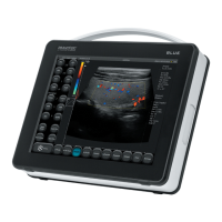
BLUE is a modern device which can be powered from a built-in battery pack or directly from the mains (110-
230 V). The ultrasound scanner is built based on a small-size computer system. The special features of the de-
vice are: touch operation, small size and weight as well as a strong aluminium casing which hides technically
advanced electronics which guarantees top quality imaging.
BLUE is a unique diagnostic device. Its small size and a built-in battery power supply make this ultrasound
scanner a modern and ergonomic tool. It can be used as a desktop or portable device. It has a at high quality
LCD LED monitor which guarantees bright and high resolution image, which makes the work easier in almost
any conditions.
BLUE is an ultrasound scanner designed for diagnostic purposes, to monitor the progression of the disease and
to evaluate the condition of the organs. Its main applications are:
- abdominal cavity,
- gynaecology and obstetrics,
- vascular ow,
- musculoskeletal system,
- heart,
- lungs imaging.
Operator can use black and white real time imaging modes as well as Color, Power and Pulse Wave Doppler.


 To Next Page