Bladder Volume Tester User’s Manual V1.02
- 17 -
Chapter Ten Bladder volume measurement
10.1 Scanning and positioning bladder
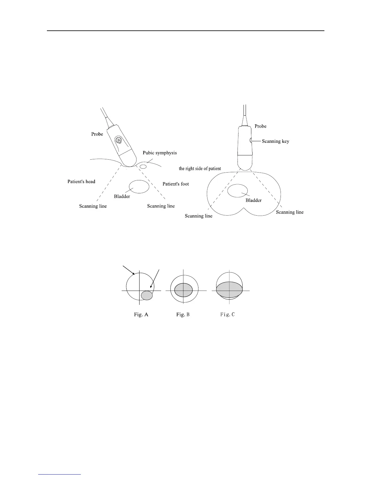
The correct positioning of bladder is the basis of accurate measurement of bladder volume. Bladder is
located in the lower abdomen, below the pubic symphysis. Before the examination, apply ultrasound
coupling gel on the subject, place the probe in accordance with the position of probe shown in the figure
below, note that the direction of scanning key on the probe toward the subject’s head.
Fig. Probe position
To determine the correct measurement position, the right side of touch screen displays the bladder
projection. If the projection was nearly round, basically in the center and not beyond the scanning border
(shown in Fig B), it indicates that the position of probe is correct and the volume is valid; otherwise it
should adjust the position of probe and re-measurement.
Fig. Projection position sketch map
Figure A shows: bladder projection obviously deviates from the centerline of the test, which is located
in the lower right of the centerline of the probe. The measured data is not accurate, it need to adjust the
angle and position of probe, and re-measurement.
Figure B shows: bladder projection basically locates in the center of test area, which was
approximately round and does not exceed the scanning boarder. The bladder volume is valid.
Figure C shows: bladder projection is beyond the border, the test data is small, it need to adjust the
angle and position of probe, and re-measurement.
The system will automatically identify the border of bladder and calculate the cross-sectional area and
volume. The green trace in the image is the border of bladder.
10.2 Operation processes
1. The subject was held in a supine position, so that the abdominal muscles to relax. First find
the pubis, and then apply an amount of ultrasound coupling gel on the pubic above 3cm from the
center of the abdomen (air bubbles as few as possible);