Chapter 2: System Overview
Page 10
• Protect the membrane from light until it has been imaged.
• Keep the membrane wet if it is to be stripped and re-used. This can easily be accomplished by wrapping the mem-
brane in plastic wrap.
• For infrared Western blots, store dry or in PBS buffer at 4°C.
• Do not dry chemiluminescent membranes for imaging.
• Use clean containers to avoid cross-contamination and reduce background.
• Multiple membranes can be washed together, provided there is ample volume so each membrane moves freely.
• The NIR fluorescent signal on the membrane will remain stable for several months or longer on blots stored dry and
protected from light and air.
Using Gels
DNA gels that are stained with ethidium bromide or other comparable stains (such as SYBR
®
Safe) can be imaged in the
600 nm channel. A detailed technical note on imaging DNA gels can be found in the Odyssey
®
Fc Application Protocols
manual. Coomassie-stained gels can also be imaged since Coomassie Blue dye can be seen clearly in the 700 nm chan-
nel, and faintly in the 800 nm channel. To image a gel, observe the following guidelines:
• Thoroughly rinse the gel with destaining solution or water to remove dye particulates.
• When placing the gel in the imaging tray, place the top toward the back of the instrument.
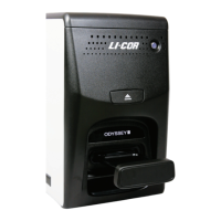
Optical System Description
The Odyssey Fc Imager is designed to image Western blots, membranes, or gels using visible, near-infrared or chemilu-
minescent methods. Before image acquisition starts, the imaging tray containing a membrane or gel is placed in the
Odyssey Fc imaging drawer and the drawer is closed. A safety interlock prevents operation of the laser illumination
module while the imaging drawer is open. Image acquisition is started via Image Studio
software.
The Odyssey Fc has four detection channels: two are infrared channels with 685-690 and 785 nm lasers, one is a 600 nm
channel with a light source for excitation, and the fourth is for detecting visible wavelength emissions from chemilumi-
nescent substrates. The channels used during acquisition are chosen in Image Studio software.
The Odyssey Fc laser module contains a 685-690 nm and 785 nm laser. During image acquisition, each laser source is
turned on, followed by image acquisition with the LI-COR
®
CCD camera. For infrared imaging, a typical image acqui-
sition consists of images for two fluorescent probes in the 700 and 800 nm channels (assuming both channels are enabled).
Image acquisitions from chemiluminescent substrates use just the chemiluminescence channel and imaging is done at
visible wavelengths. Note, however, that fluorescent markers can be imaged separately in the 700 nm channel and
combined with the image from the chemiluminescence channel. Coomassie stains can also be imaged in the 700 nm
channel. DNA gels stained with ethidium bromide or other comparable stains (such as SYBR Safe) can be imaged in the
600 nm channel.
The quality of Odyssey Fc images is enhanced by a patented filtering system that dramatically reduces noise before
detection by the CCD detector. Infrared signal detection has also been optimized for LI-COR
®
IRDye
®
near-infrared dyes,
which eliminates the need for filter selection by the user before imaging. The unique imaging technology, FieldBrite
TM
XT
2
, used in the Odyssey Fc Imager acquires images without saturated pixels on the first attempt with no user adjust-
ments. More than six logs (22 bits) of dynamic range are available for each image.