Do you have a question about the Mindray MX8 and is the answer not in the manual?
Lists situations not covered by warranty, such as improper use, man-made failure, or force majeure.
Provides contact information for Mindray and Mindray DS USA for customer support.
Defines signal words like DANGER, WARNING, CAUTION, NOTE, and TIP used in the manual.
Explains the meaning of various safety symbols used throughout the manual.
Details essential precautions for ensuring patient and operator safety during system use.
Provides a warning about potential allergic reactions to latex in system components.
Describes the system's intended applications for various patient types and exams.
Lists contraindications for system use, including FDA and Canada region specifics.
Details the system's safety classifications regarding electric shock and water ingress.
Outlines key technical specifications like imaging modes, power, and environmental conditions.
Compares features and availability of different system models (e.g., MX7, Zeus, MX8).
Lists standard configurations and available optional items for the system.
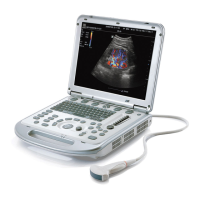
Provides an overview and description of the system's main components and ports.
Details the specifications, environmental conditions, and installation of the U-Bank.
Explains the ECG module's connections and usage for physiological signal monitoring.
Describes the functions and layout of the system's control panel with trackball/trackpad.
Illustrates the main menu screen and describes the function of each icon.
Details the different areas and elements displayed on the system's monitor screen.
Covers fundamental operations like dialog box interaction and touch screen usage.
Explains the meaning and purpose of various warning labels found on the system.
Lists and explains the various symbols used on the system's interface and hardware.
Provides instructions and safety precautions for moving and positioning the system.
Details the procedures for connecting the system to external power or using batteries.
Describes the correct procedures for powering the system on, off, and managing user login.
Explains how to adjust monitor brightness and contrast for optimal image quality.
Provides instructions for safely connecting and disconnecting ultrasound probes.
Details how to connect and manage USB devices like drives and printers.
Explains how to connect the foot switch for hands-free operation control.
Guides the user through connecting and installing different types of printers.
Configures system-wide settings including region, general options, and presets.
Allows users to assign available exam modes for specific probes.
Enables customization of measurement tools for general and application-specific menus.
Allows users to preset custom comments and define comment groups for exams.
Facilitates customization of protocols and views for automated workflows.
Covers the setup and configuration for Stress Echo protocols and editing.
Configures DICOM and HL7 settings for data transfer and communication.
Manages network settings, including transmission encryption and preset services.
Configures settings related to printer setup and image printing parameters.
Provides functions for system maintenance, data backup, and troubleshooting.
Covers system security features like drive encryption, anti-virus, and transmission encryption.
Displays system software version and other device information.
Covers entering and managing patient information for new and existing exams.
Details how to activate exams completed within 24 hours or continue paused exams.
Provides instructions for pausing or ending an ongoing patient examination.
Describes the basic B mode imaging, scanning, and parameter adjustments.
Explains Color mode for detecting blood flow, including scanning and parameter adjustments.
Details Power mode for non-directional blood flow display and its parameters.
Describes M mode for observing tissue motion, including scanning and parameter adjustments.
Explains Color M mode for cardiac motion state and its parameters.
Covers Anatomical M mode for flexible M-mark line placement and its variations.
Details Pulsed Wave (PW) and Continuous Wave (CW) Doppler modes for velocity analysis.
Explains Tissue Doppler Imaging (TDI) modes for tissue motion and velocity analysis.
Describes iScape panoramic imaging for extended field of view and its procedures.
Covers R-VQS for arterial stiffness quantification, including parameters and procedures.
Explains Smart B-line for detecting lung B lines in B mode and its procedures.
Details Smart VTI for calculating cardiac output and evaluating cardiac function.
Explains Smart IVC for measuring IVC diameter and change rate in B mode.
Covers RIMT for detecting coronary artery disease and intima thickness.
Describes Tissue Tracking QA for myocardial movement evaluation and quantitative analysis.
Explains iWorks for automating workflow, speeding up exams, and reducing keystrokes.
Details how to quickly save image parameters and settings for probes and modes.
Introduces 3D imaging and its advantages over 2D imaging for structure visualization.
Provides important notes and tips for using Smart 3D imaging effectively.
Guides the user through the basic steps for performing strain elastography scans.
Details image parameters for optimizing Elasto images, including smooth, opacity, and invert.
Describes how to perform measurements related to mass analysis in elastography.
Provides step-by-step instructions for performing contrast imaging procedures.
Details the procedure for Left Ventricular Opacification using contrast agents.
Explains Low MI Contrast Imaging for myocardial analysis using contrast agents.
Describes image parameters specific to contrast imaging, including timer settings.
Covers saving live capture and cine images during contrast imaging.
Explains Micro Flow Enhancement (MFE) for visualizing tiny vessel structures.
Details Contrast Imaging QA for time-intensity analysis and perfusion quantification.
Guides on connecting ECG electrodes and system setup for physiological signal monitoring.
Describes how to acquire and display respiratory wave signals.
Explains how to review ECG waveforms and linked image data.
Provides descriptions for various physio parameters like ECG source, gain, and speed.
Details the steps for acquiring Stress Echo loops, including protocol selection and ROI setup.
Explains how to select the best loops for analysis in review and wall motion scoring modes.
Covers reviewing and scoring cardiac wall motion abnormalities in Stress Echo exams.
Describes how Stress Echo data, including loops and scores, is saved.
Provides instructions on how to exit the Stress Echo feature.
Explains how to perform measurements and generate reports for Stress Echo exams.
Covers system support for dual-split and quad-split display formats.
Details how to zoom in on images using Spot Zoom, Pan Zoom, and iZoom functions.
Explains how to freeze and unfreeze scanning images and switch modes when frozen.
Guides on reviewing and editing images prior to freezing, supporting manual and auto review.
Describes how to compare images from the same or different exams.
Details the process of saving live capture and frozen images or cine clips.
Explains how to set the duration for saving cine clips in live and frozen modes.
Covers general and application measurements, including accuracy specifications.
Details how to add, move, edit, and delete text, arrows, and traces as annotations.
Explains how to add, move, and delete body marks for indicating patient or probe position.
Covers storing, managing, and exporting image files using various media and formats.
Details procedures for managing reports, including importing, exporting, and sending.
Explains how to search, view, backup, send, restore, and delete patient data in iStation.
Describes how to recover deleted patient data from the recycle bin.
Covers saving image files and measurement reports to a remote PC server via iStorage.
Provides information on printer connection and settings for image and report printing.
Details the process of backing up files to CD/DVD using the system's drive.
Manages system tasks such as storage, printing, and DICOM service settings.
Used for sending images or structured reports to a DICOM storage server.
Used for sending images to a DICOM print server for printing.
Enables querying or importing patient data from a Worklist server.
Sends exam state information to the configured server for progress tracking.
Confirms successful storage of images or reports on the DICOM storage server.
Allows querying and retrieving patient exam records from a designated server.
Details saving and reviewing patient data in DCM format on external media.
Describes procedures for sending structured reports, including storage options.
Lists available probes, their region applied, intended use, imaging modes, and figures.
Covers warnings, procedures, and cleaning/sterilization for biopsy operations.
Explains the Middle Line function for locating lithotripsy focus points.
Details the eSpacial Navi function for ultrasound needle guidance.
Provides instructions on how to start and save video/audio recordings.
Covers exporting recorded images saved in the local disk.
Explains how to replay recorded videos and audio on a PC or the system.
Outlines routine daily maintenance tasks, including cleaning and checks.
Provides a table of common system failures, their causes, and troubleshooting measures.
Details operation modes, setup, and scanning techniques for 1-D barcode readers.
Describes the overview, setup, and settings for the 2-D barcode reader.
Covers supported JADAK models and their configuration procedures.
Provides instructions for cleaning the barcode reader's exit window.
Lists the default parameters for LS2208 and DS4308 barcode readers.
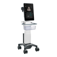
Lists the supplied accessories for the MT3 Trolley.
Provides an overview of the MT3 and MT2 Trolley units and their components.
Details inspection criteria for the power plug, body, and strain relief.
Covers visual and contextual inspections of the device enclosure and accessories.
Ensures that manufacturer and warning labels are present and legible.
Specifies the procedure and limits for testing protective earth resistance.
Describes the procedure and limits for performing Earth Leakage tests.
Guides on connecting to and using the wireless LAN function.
Details how to configure IP settings for the system's network connection.
Provides information on setting up EAP network connections.
Explains how to use iScanHelper for scanning reference and obtaining guidance.
Describes how to use iScanHelper for learning scanning techniques and practicing views.
Covers the basic screen layout and operations of the iScanHelper tool.
Describes demonstration items as image files supported by the system.
Explains the two types of catalogs: Demo Catalog and Customize Catalog.
Provides instructions for transferring files between mobile hard disk and ultrasound system.
Describes how the system automatically plays image files in the list.
Allows choosing to repeat demonstration or exit after completion.
Allows switching to B Mode using a vocal command.
Allows freezing the image using a vocal command.
Allows turning the full image mode on or off using a vocal command.
Allows switching exam mode to Adult Abdomen via vocal command.
Allows switching exam mode to Adult Cardiac via vocal command.
Allows opening the patient info dialog box via vocal command.
Discusses potential bioeffects of diagnostic ultrasound and safety recognition.
Emphasizes the importance of using ultrasound prudently for patient benefit.
Explains the ALARA principle for controlling total energy and output intensity.
Explains Mechanical Index (MI) and Thermal Index (TI) as bioeffect indicators.
Covers adjusting acoustic power percentage and its default settings.
Categorizes system controls affecting ultrasound output and direct/indirect controls.
Details derated ultrasonic output parameters and their operational limits.
Lists the total estimated measurement uncertainties for various acoustic quantities.
Provides references for acoustic power and safety standards and guidelines.
Provides guidance on electromagnetic emissions compliance and environment.
Provides guidance on electromagnetic immunity compliance and environment.
| Touch Screen | Yes |
|---|---|
| Display | LED |
| Display Size | 15.6 inches |
| Battery Life | Up to 3 hours |
| Imaging Modes | B/M/Color/Power/PW/CW |
| Operating Modes | General Imaging, Cardiology, Vascular, Abdominal, Obstetrics, Gynecology, Small Parts, Musculoskeletal, Urology, Pediatric |
| Connectivity | DICOM, Ethernet, WiFi |
| Ports | USB, HDMI, VGA, Ethernet, Audio In/Out |
| Operating System | Windows |