EasyManua.ls Logo

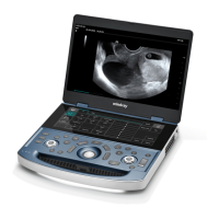
Mindray ME7

Mindray ME7
338 pages
To Next Page IconTo Next Page
Loading...
MX7/MX7 Exp/MX7T/MX7S/MX7 Pro/
Vaus7/Zeus7/Zeus/ME7/ME7 Exp/Anesus
ME7/Anesus ME7 Pro/Anesus ME7S/
Anesus ME7T/Ana/Anesus Vaus/Anesus
Zeus/Crius ME7P/Crius ME7W/Anesus
ME7P/Anesus ME7W/Emerus ME7P/
Emerus ME7W/MX7P/MX7W
Diagnostic Ultrasound System
Operators Manual
[Basic Volume]

Table of Contents

Other manuals for Mindray ME7

Questions and Answers:

Mindray ME7 Specifications

General IconGeneral
TypePortable Ultrasound System
Touch ScreenYes
Image ModesColor Doppler, Power Doppler
Operating ModesM-Mode
Transducer TypesConvex, Linear, Phased Array
ConnectivityWi-Fi, Ethernet, USB, HDMI, DICOM 3.0

Summary

1 Safety Precautions

1.1 Meaning of Safety Symbols

Explains the meaning of various safety symbols used throughout the manual.

1.2 Safety Precautions

Provides essential precautions for ensuring patient and operator safety during system use.

1.3 Latex Alert

Warns about potential allergic reactions to latex in probe sheaths and components.

2 System Overview

2.1 Intended Use

Details the intended applications and body parts for the ultrasound system's use.

2.3 Safety Classifications

Outlines the system's safety classifications regarding electric shock, water ingress, and anesthetic mixtures.

2.4 Product Specifications

Lists key specifications including imaging modes, power supply, and environmental conditions.

2.6 System Configuration

Describes the standard configuration and available options for the system.

3 System Preparation

3.1 MovePosition the System

Provides instructions on safely moving and positioning the ultrasound system.

3.2 Connecting the Power Cord

Details the process of connecting the system to external power or using batteries.

3.3 Power ONOFF

Explains the correct procedures for powering the system on and off.

3.5 ConnectingDisconnecting a Probe

Provides instructions on safely connecting and disconnecting ultrasound probes.

4 Setup

4.1 System Preset

Covers system presets for region, language, time, and general settings.

4.1.7 Key Configuration

Explains how to assign functions to foot switches and user-defined keys.

4.1.10 Access Control

Details user account management, privileges, and password policies.

4.3 Measurement Preset

Allows users to preset measurement tools and parameters for different exam modes.

5 Exam Preparation

5.1 Patient Information

Covers entering and managing new patient information for exams.

5.2 Activate& Continue an Exam

Explains how to activate completed or continue paused exams.

6 Image Acquisition

6.1 B Mode

Describes the basic B mode imaging, scanning, and parameters.

6.2 Color Mode

Details the Color mode for detecting blood flow direction and speed.

6.3 Power Mode

Explains the Power mode for non-directional blood flow display.

6.8 PWCW Mode

Covers Pulsed Wave and Continuous Wave Doppler modes for flow analysis.

7 Smart 3 D

7.1 Overview

Introduces 3D imaging concepts and terminology.

7.1.2 ROI and VOI

Explains how to define Region of Interest (ROI) and Volume of Interest (VOI).

7.2.1 Smart 3 D Image Acquisition

Provides procedures for acquiring 3D images using Smart 3D.

8 Strain Elastography

8.1 Basic Procedure for Strain Elastography

Details the steps for performing strain elastography scans.

8.2 Image Parameters

Lists and explains the adjustable image parameters for elastography.

9 Contrast Imaging

9.1 Basic Procedures for Contrast Imaging

Guides on performing contrast imaging with ultrasound contrast agents.

9.4 Image Parameters

Details image parameters specific to contrast imaging.

10 Physiological Unit Signal

10.1 ECG

Explains the use of ECG signals for ultrasound imaging.

10.3 ECG Review

Describes the principle and process of reviewing ECG data.

11 Stress Echo

11.1 Stress Echo Acquisition Procedure

Details the procedure for acquiring stress echo loops.

11.2 Selecting Preferred Stress Echo Loops (Select Mode)

Guides on selecting preferred loops for analysis and comparison.

11.3.2 Wall Motion Scoring

Explains how to assign wall motion scores to cardiac segments.

12 Display & Cine Review

12.1 Splitting Display

Covers dual-split and quad-split display formats for image comparison.

12.3 FreezeUnfreeze the Image

Explains how to freeze and unfreeze scanning images.

12.4 Cine Review

Details the process of reviewing and editing captured cine images.

13 Measurement, Comments and Body Mark

13.1 Measurement

Covers general and application measurements on various image types.

13.2 Comments

Explains how to add text, arrows, and traces as comments to images.

13.3 Body Mark

Describes how to add and manage body marks for indicating probe position.

14 Patient Data Management

14.1 Image File Management

Covers storing, reviewing, and analyzing image files on various media.

14.2 Report Management

Details importing, exporting, and sending patient reports.

14.3 Patient Data Management (iStation)

Explains how to search, view, and manage patient data using iStation.

15 DICOMHL7

15.1 DICOM Storage

Covers sending images and reports to DICOM storage servers.

15.6 QueryRetrieve

Explains how to query and retrieve patient exam records from a server.

16 Probes and Biopsy

16.1 Probes

Lists available probes, their applications, and remarks.

16.1.5 Probes Cleaning and DisinfectionSterilization

Provides detailed procedures for cleaning and disinfecting probes.

16.2 Biopsy Guide

Covers warnings and procedures related to biopsy guidance.

16.2.2 Needle-Guided Bracket Inspection and Installation

Explains how to inspect and install needle-guided brackets.

16.2.4 Starting the biopsy procedure

Details the crucial steps and safety precautions for starting a biopsy.

17 DVR Recording

17.1 Start Recording

Provides instructions on starting and saving video recordings.

18 System Maintenance

18.1 Daily Maintenance

Covers daily tasks for system cleaning and checks.

18.2 Troubleshooting

Lists common system malfunctions and their solutions.

A Barcode Reader

A.1 1-D Barcode Reader

Details the operation modes and setup for 1-D barcode readers.

A.2 2 D Barcode Reader (Take DS4308 as an example)

Explains the setup and operation of the 2D barcode reader.

B Trolley and Accessories

B.1 Option Accessories

Lists optional accessories for the ultrasound system.

B.2 Introduction of Each Unit

Provides an overview and description of the trolley's components.

C Electrical Safety Inspection

C.1 Power Cord Plug

Details the inspection criteria for the power cord and plug.

C.4 Protective Earth Resistance

Outlines the procedure and limits for testing protective earth resistance.

C.5 Earth Leakage Test

Describes the Earth Leakage test procedure and conditions.

D Wireless LAN

D.1 Use the Wireless feature

Guides on connecting to and using the wireless LAN feature.

E iScanHelper

E.1 Use iScanHelper for Reference

Explains how to use iScanHelper for scanning reference and guidance.

E.3 Basic Screen and Operation

Describes the basic screen layout and operations within iScanHelper.

G List of Vocal Commands

H Acoustic Output

H.3 ALARA Principle (As Low As Reasonably Achievable)

Explains the ALARA principle for controlling ultrasound exposure.

H.5 Acoustic Power Setting

Details how to adjust acoustic power and its default settings.

H.6 Acoustic Power Control

Describes system controls that affect ultrasound output.

I EMC Guidance and Manufacturers Declaration

Related product manuals