Do you have a question about the Mindray Z50BW and is the answer not in the manual?
| Brand | Mindray |
|---|---|
| Model | Z50BW |
| Category | Medical Equipment |
| Language | English |
Describes the purpose and content of the operator's manual.
Explains symbols and conventions used throughout the manual.
Classifies the equipment based on protection against electric shock and water ingress.
Defines signal words like DANGER, WARNING, CAUTION, and NOTE used for safety.
Explains various safety symbols used in the manual and on the system.
Provides detailed safety precautions for patient and operator.
Provides a warning regarding potential allergic reactions to latex.
Explains the meaning and purpose of warning labels on the system.
Specifies the intended applications and clinical areas for the system.
Lists conditions or uses for which the system is not intended.
Details technical specifications including imaging modes, power, and environmental conditions.
Outlines the main unit, software, and accessories included in the standard configuration.
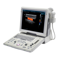
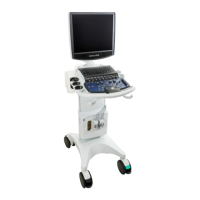
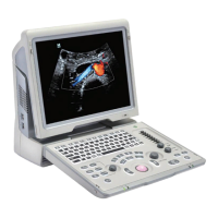
Describes the main components and their functions, including panels and ports.
Explains system symbols and their meanings for user reference.
Instructions for safely moving and positioning the ultrasound system.
Covers connecting the system to external power or using battery backup.
Procedures for safely turning the system on and off.
Steps for attaching and detaching ultrasound probes.
Instructions for using USB devices for data transfer.
Details on connecting and configuring graph/text printers.
Overview of the system's main screen layout and basic operations.
Describes how to begin a new patient exam or continue an existing one.
Covers entering and managing patient demographic and exam data.
Guides the selection of appropriate exam modes and ultrasound probes.
Procedures for reactivating or continuing a previously started exam.
Instructions on how to pause or conclude an ultrasound examination.
Fundamental adjustments for optimizing image brightness, contrast, and grayscale.
Detailed optimization of the B Mode imaging parameters.
Optimization procedures and parameters specific to M Mode imaging.
Adjustments for optimizing Color Doppler imaging parameters.
Optimization techniques for Power Mode imaging.
Optimization of Pulsed Wave Doppler imaging parameters.
Using the Free Xros M feature for anatomical M Mode imaging.
Optimization of Tissue Doppler Imaging modes.
Procedures and optimizations for iScape panoramic imaging.
Overview and optimization of 3D and 4D imaging capabilities.
Options for splitting the display and magnifying images.
How to review and analyze recorded cine clips.
Functionality for comparing multiple images.
Managing and clearing cine memory.
Setting and managing image presets.
General steps for entering and exiting measurement functions.
Performing standard measurements in 2D, M, and Doppler modes.
Using measurement tools specific to clinical applications.
Information on the accuracy of various measurement tools.
Procedures for adding, editing, moving, and deleting text comments.
Procedures for adding, moving, and deleting body mark pictograms.
Entering and managing patient demographic information.
Storing, organizing, and reviewing image files.
Managing and handling patient reports.
Using the iStation system for comprehensive patient data management.
Procedures for backing up and erasing data using a DVD drive.
Monitoring and managing system tasks.
Setting up and managing user access and security.
Initial setup and configuration for DICOM network and services.
Checking and troubleshooting DICOM network connections.
Setting up and managing DICOM services like storage, print, and worklist.
Saving patient data to external media in DICOM format.
Handling DICOM structured reports.
Monitoring and managing DICOM tasks.
Configuring system-wide preferences like region, general, image, and application settings.
Setting up exam-specific presets for probes and modes.
Customizing measurement settings.
Configuring comment libraries and presets.
Customizing body mark presets for exam types.
Configuring printer settings and services.
Setting up network connections and iStorage.
System maintenance procedures and options.
Viewing system details like software and hardware versions.
Details on probe models, parts, orientation, and handling.
Guidance and procedures for performing ultrasound-guided biopsies.
Information on the Lithotrity function for treatment guidance.
General information about the system's battery.
Safety precautions for handling and using the battery.
Procedures for installing and removing the battery pack.
Understanding the battery status indicator on the screen.
Methods for checking the battery's performance over time.
Discusses potential biological effects of ultrasound exposure.
Guidelines for using ultrasound prudently to benefit the patient.
Explains the ALARA principle for controlling ultrasound exposure.
Details on Mechanical Index (MI) and Thermal Index (TI) parameters.
How to adjust and control the system's acoustic power output.
Overview of controls that affect ultrasound output.
Information on derated output parameters and limits.
Details on the uncertainty associated with acoustic output measurements.
List of references for further information on acoustic power and safety.
Routine daily cleaning and checks for the system.
Periodic checks performed by authorized service personnel.
Information on replaceable consumables and parts.
Guidance for diagnosing and resolving system malfunctions.
Inspection criteria for the power cord and plug.
Visual and contextual inspection of the device enclosure and accessories.
Testing the protective earth resistance of the system.
Performing earth leakage tests on the device.
Testing for leakage current from the device enclosure.
Measuring patient leakage current.
Testing leakage current on applied parts connected to mains.
Measuring patient auxiliary currents.
Overview and setup of the 1D barcode reader.
Default settings for the barcode reader's parameters.
Maintenance procedures for the barcode reader's window.
Explains the objective and benefits of the iWorks workflow automation.
Step-by-step guide for using the iWorks protocol.
Description of the iWorks screen layout and elements.
Operations related to selecting and managing views within iWorks.
Safety guidelines for using the printer adapter.
List of printers supported by the adapter.