Do you have a question about the Siemens Acuson S2000 and is the answer not in the manual?
Provides an overview of the ACUSON S Family ultrasound systems and their capabilities.
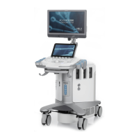
Details the components and layout of the ultrasound system.
Describes the physical controls and layout of the ultrasound system's control panel.
Specifies the intended applications and usage guidelines for the ultrasound system.
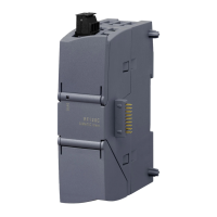
Lists compatible transducers and their intended applications for the ultrasound system.
Describes the layout and components of the ultrasound system's image screen.
Provides essential safety information regarding electrical connections and precautions.
Details daily checks, maintenance, and cleaning procedures for the ultrasound system.
Provides guidelines on cleaning, disinfecting, and handling transducers.
Explains how to care for transducer accessories such as sheaths and needle guides.
Covers product recycling, disposal, and hazardous substances.
Provides instructions for battery recycling and disposal according to regulations.
Covers initial setup procedures, including safety warnings for system handling.
Provides instructions and safety precautions for moving the ultrasound system.
Details the procedures for powering on and starting the ultrasound system.
Explains how to connect and supply power to the ultrasound system.
Provides instructions for connecting and disconnecting transducers to the system.
Details how to connect system accessories like footswitches and physio cables.
Explains how to establish wired and wireless network connections for the system.
Covers features for protecting patient confidentiality and restricting user access.
Describes the various ports and connections on the input/output panel.
Guidelines for connecting peripheral equipment to the ultrasound system.
Details adjustments for monitor height, swivel, and control panel positioning.
Explains how to secure the ultrasound system and manage user access.
Covers procedures for registering patients and managing patient data.
Provides an overview of imaging modes and on-screen display elements.
Details how to use the measurement function for various exam types.
Explains how to review, manage, and print images and clips.
Describes how to create and view thumbnail representations of images and clips.
Instructions for using the DVR to record and play back patient studies.
Provides general information, application guidelines, and disposal instructions for transducer sheaths.
Describes the gel pad, its preparation for use, and storage.
Information on needle guide brackets, including attachment and care procedures.
Explains the biopsy function, activating on-screen guidelines, and system safeguards.
Details the process for verifying the needle path using on-screen guidelines.
Provides an introduction to transesophageal transducers and TEE procedures.
Describes the V5Ms transducer, its modes of operation, and controls.
Details the V7M transducer, its modes of operation, and controls.
Covers cleaning, disinfection, storage, and handling of the transesophageal transducer.
Outlines preventive measures to ensure patient safety and equipment integrity.
Information about the 9EVF4 transducer, its markings, and imaging capabilities.
Describes the markings on the 9EVF4 transducer, including the orientation indicator.
Details how to use the 9EVF4 transducer for imaging and volume acquisition.
Provides technical specifications for the 9EVF4 transducer.
Describes the physiologic function module, including ECG features.
Provides step-by-step instructions for activating the ECG feature.
Lists common ECG issues and their possible causes and recommended solutions.
Provides an overview of eSieFusion imaging, its features, and compatibility.
Instructions on how to activate and deactivate the eSieFusion feature.
Details the process of importing and loading reference data for eSieFusion.
Explains how to align reference data with ultrasound images using alignment tools.
Provides guidance on resolving common issues with the eSieFusion tracking system.
Covers connecting and setting up the eSieFusion tracking system components.
Guidelines for maintaining, cleaning, and caring for eSieFusion equipment.
Explains the ARFI technology used in Virtual Touch features.
Describes how to qualitatively visualize tissue stiffness using Virtual Touch imaging.
Explains how to quantitatively measure tissue shear velocity and elasticity.
Details how to quantitatively depict tissue stiffness and measure shear wave velocity.
Provides tips for scanning techniques to improve Virtual Touch IQ results.
Lists the standard features of the ACUSON ultrasound systems.
Describes the features and adjustments of the operator control panel.
Details the system operating software, including language and customization options.
Provides specifications and adjustments for the high-resolution flat panel display.
Describes the system's mobility features, including braking and steering.
Highlights the technologies and design attributes of the ultrasound transducers.
Lists the available USB, Ethernet, and other connection ports on the system.
Details the single and combined operating modes supported by the system.
Describes the general imaging features available for 2D-mode.
Details the features and measurements available for Pulsed Wave Doppler.
Describes the features and selections for Color Doppler Velocity imaging.
Details the features and selections for Color Doppler Energy imaging.
Lists the features and controls available for M-Mode imaging.
Lists Siemens-authorized accessories and options for the ultrasound system.
Information on available language options for the operating system.
Details various optional features and technologies available for the ultrasound system.
Describes the advanced SieClear option for enhanced image viewing.
Information on the eSieFusion imaging feature and its supported components.
Provides qualitative representation of tissue stiffness using eSie Touch.
Details the Virtual Touch Imaging option and its compatible transducers.
Describes the Virtual Touch Quantification option and its compatible transducers.
Information on the Virtual Touch IQ option for quantitative stiffness measurements.
Details the contrast agent imaging option, including acquisition techniques.
Explains the syngo Auto Left Heart analysis option for cardiac assessment.
Describes the syngo Velocity Vector Imaging option for motion analysis.
Lists the available digital documentation devices and consumables.
Covers general and application-specific measurements and report generation.
Details the general functions for measurements and reports.
Lists application-specific measurements and calculations for various exam types.
Provides accuracy and range specifications for clinical measurements.
Outlines system requirements including power supply and combinations with other equipment.
Details system classifications regarding electrical shock and ingress protection.
Lists the standards and compliance relevant to the ultrasound system.
Describes the control panel, its lighting, and audio settings.
Details the touch screen layout and symbols used for selections.
Explains the use and functions of the trackball and soft keys.
Describes controls for activating and adjusting imaging modes like 2D, Color, Doppler, M-Mode.
Details controls for optimizing image parameters such as depth, focus, and scale.
Explains controls for adding annotations and performing measurements.
Describes controls for freezing images and archiving data.
Details specialty controls like UD, Pointer, and Forward/Backward.
Explains advanced controls such as 3D/4D, Elasticity, and Contrast.
Covers controls for patient management, review, report generation, and exam end.
Describes the retractable and touch screen alphanumeric keyboards.
Details special keys available on both keyboards for various functions.
Describes the control panel layout, keys, and lighting for non-touch screen systems.
Explains the status indicators based on control lighting colors.
Details the trackball and soft key functions and their active tasks.
Explains the function assigned to the UPDATE key for various active functions.
Describes how the SELECT key interacts with controls for imaging, measurement, and review.
Explains how the PRIORITY control cycles through imaging modes and tools.
Describes how the NEXT control cycles through imaging mode tools.
Details the GAIN FREEZE/CINE control for adjusting gain and managing images/clips.
Explains the controls for activating and adjusting imaging modes like 2D, Color, and M-Mode.
Describes controls for optimizing image parameters like depth, focus, and scale.
Explains how to adjust transmit power for the active priority mode.
Details controls for CADENCE, DUAL, and REVIEW functions.
Describes controls for DVR, CLIP STORE, PRINT, and STORE functions.
Explains functions based on current system state or operating mode.
Details the special characters available on the alphanumeric keyboard.
Lists and describes the function keys available on the control panel.
Provides an overview of on-screen controls and how they are displayed.
Describes the toolbar selections for accessing functions like Patient Browser and System Config.
Explains the thumbnail panel for viewing stored clips, volumes, and images.
Details how to adjust control panel lighting and audio settings.
Describes mode-dependent parameters for adjusting imaging settings.
Explains controls for customizing and managing eSieScan workflow protocols.
Details parameter selections for 2D-mode imaging.
Describes parameter selections for M-mode imaging.
Details parameter selections for Color Flow Imaging (CDV, CDE, DTI).
Explains selections for Doppler parameters like filter, sweep speed, and maps.
Describes settings for storing and reviewing clips, including capture definition.
Details selections for adding text, body markers, arrows, and setting home position.
Explains basic measurement tools like Delete, Undo, and APAD/TAD.
Details measurement tools for 2D-mode imaging, including distance and ellipse.
Describes measurement tools for Doppler imaging, including velocity and heart rate.
Lists measurement tools for M-mode imaging, such as distance and slope.
Explains controls for navigating and managing the patient report.
Details basic cardiac measurement tools and cardiac report controls.
Explains DVR menu selections and status indicators for recording.
Describes controls for reviewing images, clips, and protocols.
Covers controls for previewing, customizing, and printing images.
Describes the patient browser screen, icon selections, and menu options.
Details menu selections for patient data management and transfer.
Explains selections within the patient menu for data management.
Covers selections for transferring patient data to local or external storage.
Details selections for editing patient data, including cut, paste, and protect.
Explains selections for viewing patient data in different layouts.
Describes filter selections for displaying patient data based on criteria.
Explains sorting options for patient data across different locations.
Provides a description of controls for the eSieFusion imaging feature.
Details selections for eSie Touch elasticity imaging.
Describes tools for making elasticity measurements, such as shadow and strain ratio.
Provides an overview of controls for Virtual Touch imaging features.
Details controls for 3D and 4D imaging setup and editing.
Explains selections for configuring 3D and 4D imaging parameters.
Describes selections for acquiring and adjusting 3D and 4D images.
Details selections for visualizing 3D and 4D data, including render modes.
Covers editing tools for 3-Scape and fourSight imaging.
Details controls for SieScape imaging, including scan start, pause, and resize.
Describes selections for contrast agent imaging, including burst and MBD techniques.
Explains controls for reviewing reference images from other modalities.
Describes the reference images screen for selecting and viewing images.
Provides information on controls related to the physiologic function.
Details selections used within the Stress Echo feature.
Explains controls for the syngo Auto Left Heart analysis.
Describes controls for syngo Velocity Vector Imaging.
Details controls and context menus for Contrast Dynamics software.
Explains controls for the syngo Arterial Health Package (AHP).
Discusses factors affecting index display accuracy and measurement uncertainty.
Provides default MI and TI values for various transducers and modes.
Presents acoustic output data according to FDA and IEC standards.
Provides a summary table of MI or TI values exceeding 1.0 for each transducer/mode.
Defines key terms and symbols used in the acoustic output reporting tables.
Provides a legend for understanding the acoustic output reporting tables.
| Type | Ultrasound System |
|---|---|
| Manufacturer | Siemens |
| Model | Acuson S2000 |
| Power Supply | 100-240 VAC, 50/60 Hz |
| Frequency Range | 1 - 18 MHz (depending on transducer) |
| Transducers | Volume transducers |
| Imaging Modes | B-Mode, M-Mode, Color Doppler, Power Doppler, Pulsed Wave Doppler, Continuous Wave Doppler, Tissue Doppler |
| Applications | Cardiology, obstetrics, gynecology, abdominal imaging |
| Advanced Imaging Technologies | Virtual Touch |
| Portability | Mobile cart-based system |
| Connectivity | DICOM compatible, USB, network |