Do you have a question about the Siemens Acuson S3000 and is the answer not in the manual?
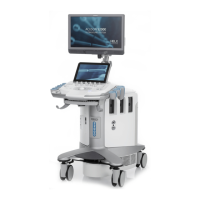
Description of the control panel layout, including keys, controls, and customizable options.
Details on intended applications and warnings for the ultrasound system.
Information on intended applications where the ultrasound system should not be used.
Guidelines for safe operation and environmental considerations for the ultrasound system.
Critical safety information regarding electrical connections, grounding, and potential hazards.
Precautions and warnings related to handling biohazardous materials and patient safety.
Information on mechanical and thermal indices and their display on the ultrasound system.
Important initial setup procedures and safety warnings before operating the system.
Instructions and precautions for safely moving the ultrasound system.
Steps for connecting the system to power and initiating the startup sequence.
Essential procedures for safely connecting and disconnecting transducers.
Features and configurations for protecting patient confidentiality and system access.
Safety and compliance requirements for connecting peripheral devices to the ultrasound system.
Procedures and system features to protect patient information from unauthorized access.
Steps required to log in to the ultrasound system when the security package is activated.
Detailed instructions for registering new patients or pre-registering scheduled patients.
Procedures for utilizing the measurement and calculation functions.
Overview of available transducer accessories and their compatibility.
Explanation of the on-screen guidelines for biopsy procedures.
Safety measures and precautions to reduce the risk of injury during biopsy procedures.
Procedures for verifying the accuracy of the needle path indicated by the on-screen guidelines.
Overview of transesophageal transducers and their use in TEE procedures.
Precautions and procedures to follow before using the transesophageal transducer.
Procedures for cleaning and disinfecting the transesophageal transducer.
Comprehensive list of preventive measures to ensure patient safety and equipment protection.
Overview of the Physio module and its ECG feature, including warnings and precautions.
Step-by-step instructions for activating the ECG feature and connecting leads.
Introduction to eSieFusion imaging, its features, and compatible equipment.
Procedures for activating and deactivating the eSieFusion feature.
Tools and procedures for aligning reference data with ultrasound images.
Common symptoms and recommended actions for troubleshooting the eSieFusion tracking system.
Procedures for connecting and setting up the tracking system components.
Instructions on using Virtual Touch imaging to qualitatively visualize tissue stiffness.
Instructions on using Virtual Touch quantification to measure tissue shear velocity or elasticity.
Information on using Virtual Touch IQ for quantitatively depicting tissue stiffness.
Overview of the selections and functions available on the touch screen.
Explanation of how SELECT and SELECT-R keys interact with controls and on-screen items.
Description of controls for activating and adjusting various imaging modes.
Controls for adding annotations and performing measurements.
Controls for managing patient, review, report, and exam workflows.
Description of the control panel for systems without a touch screen, including lighting.
Description of the trackball and soft key controls and their functions.
Description of controls for activating and adjusting various imaging modes.
Description of controls for optimizing image settings like depth, focus, and scale.
Acoustic output reporting tables for transducer models exceeding MI or TI value of 1.0.
| Type | Ultrasound System |
|---|---|
| Power Supply | 100-240 VAC, 50/60 Hz |
| Portability | Mobile |
| Connectivity | DICOM, USB, Ethernet |
| Frequency Range | 1.0 - 18 MHz (depending on the transducer) |
| Imaging Modes | B-mode, M-mode, Color Doppler |
| Probes | linear, convex, and phased array |
| Applications | Cardiology, obstetrics, gynecology, abdominal, vascular |
| Weight | Approximately 90 kg (198 lbs) |