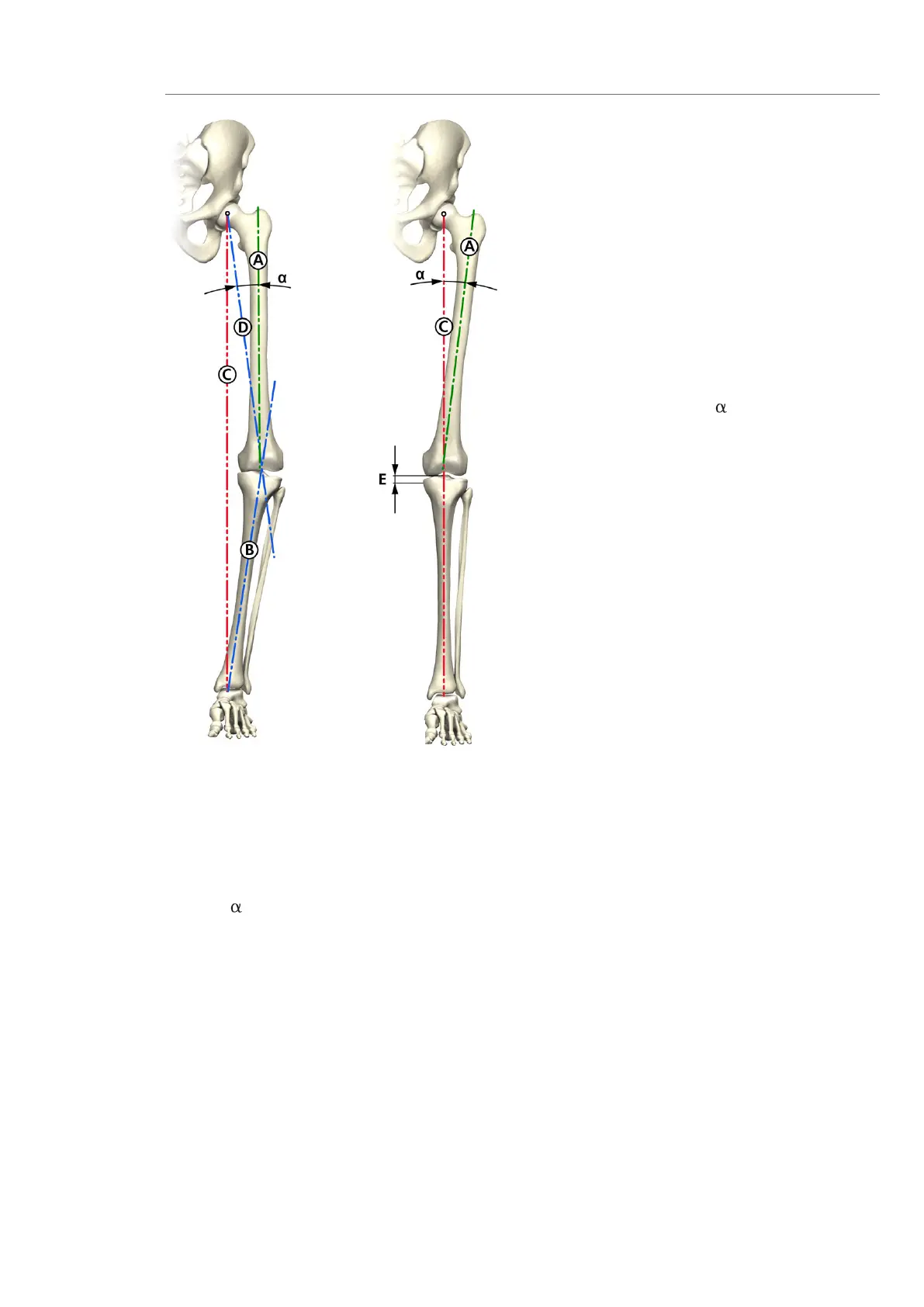
11
The following procedure is recommended
for the anterior-posterior whole leg imaging
process:
1. Femoral axis “A” (anatomical axis) is drawn
onto the radiograph.
2. A line is drawn from the femoral head to the
centre of the knee (mechanical axis “D”) on
the radiograph.
3. Angle measured between anatomical and
mechanical axes: Angle determines the
valgus angle.
4. Tibial axis “B” is drawn in and tibial resection
plane “E” is determined, in order to avoid
excessive resection, especially if defects are
present.
5. Component sizes and resection depths are
determined preoperatively, using the x-ray
templates (See page 57) in the A/P and
lateral planes.
6. Mechanical leg axis “C” should merge with
lines “D” and “B” after correction.
Planning the Surgery Using the Radiograph
A Anatomical femoral axis
B Anatomical tibial axis
C Mechanical leg axis
D Mechanical femoral axis
E Tibial resection depth (mm)
Valgus angle
Pre-op
Post-op