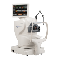
Scan Types
Macula
3D Macula – Scans the macula area 7 x 7 mm cube scan
3D Wide – Scans macula and disc combined 12 x 9 mm scan
FGA Mode – Allows precise follow up scans of Naevi and areas of interest
Dynamic Focus – Line scan allowing enhanced view of vitreous, retina and choroid in one
Radial / 5 Line Cross – Overlapping scan that penetrates cataracts or media opacities
Macula Fundus Photo – Colour, Red Free, Autofluorescence, Fluorescein Angiography
Glaucoma
3D Disc – Analysis of Retinal Nerve Fibre Layer 6 x 6 mm cube scan
3D Macula V – Analysis of Ganglion Cell Layer 7 x 7 mm cube scan
3D Wide Scans macula and disc combined 12 x 9 mm scan
Stereo Fundus – Stereo photo of optic disc
Disc Fundus Photo – Colour, Red Free, Autofluorescence, Fluorescein Angiography
Anterior
Radial Anterior 6 mm – Central Corneal Thickness measurement
Radial Anterior 16 mm – Assess larger surface area of corneal e.g scleral lens fitting
Line Anterior H or V 3 mm or 6 mm – Single Angle measurement
Line Anterior H or V 16 mm – Angle to Angle measurement
3D Anterior – 3D view of anterior angle
OCT Angiography
3 x 3, 4.5 x 4.5, 6 x 6, 9 x 9, 12 x 12 mm cube OCTA – Dye-less angiogram of structure and
flow of retinal circulation.
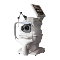
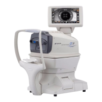
8 Topcon (Great Britain) Medical Limited | DRI OCT Triton | Quick Reference Guide | Scan Types
Scan Types
Macula
3D Macula – Scans the macula area 7 x 7 mm cube scan
3D Wide – Scans macula and disc combined 12 x 9 mm scan
FGA Mode – Allows precise follow up scans of Naevi and areas of interest
Dynamic Focus – Line scan allowing enhanced view of vitreous, retina and choroid in one
Radial / 5 Line Cross – Overlapping scan that penetrates cataracts or media opacities
Macula Fundus Photo – Colour, Red Free, Autofluorescence, Fluorescein Angiography
Glaucoma
3D Disc – Analysis of Retinal Nerve Fibre Layer 6 x 6 mm cube scan
3D Macula V – Analysis of Ganglion Cell Layer 7 x 7 mm cube scan
3D Wide Scans macula and disc combined 12 x 9 mm scan
Stereo Fundus – Stereo photo of optic disc
Disc Fundus Photo – Colour, Red Free, Autofluorescence, Fluorescein Angiography
Anterior
Radial Anterior 6 mm – Central Corneal Thickness measurement
Radial Anterior 16 mm – Assess larger surface area of corneal e.g scleral lens fitting
Line Anterior H or V 3 mm or 6 mm – Single Angle measurement
Line Anterior H or V 16 mm – Angle to Angle measurement
3D Anterior – 3D view of anterior angle
OCT Angiography
3 x 3, 4.5 x 4.5, 6 x 6, 9 x 9, 12 x 12 mm cube OCTA – Dye-less angiogram of structure and
flow of retinal circulation.
8 Topcon (Great Britain) Medical Limited |
DRI OCT Triton | Quick Reference Guide | Scan Types
8
61647 Topcon quick start guide Triton.indd 8 27/03/2019 09:56

 Loading...