7. PAN detector. This at panel detector is indicated for use in generating radiographic images of the maxillo-facial region,
more specically it is dedicated for PAN acquisitions.
8. Patient support. The patient support allows stabilising and immobilising the patient. It can be moved vertically in order to
obtain the best superimposition between the patient’s head anatomy and the height of Field of View (FOV).
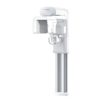
9. F group. It is the whole assembled mobile group of the device. It is the moving part of the medical device, which adapts
the acquisition geometry to the patient’s anatomy and stance (sitting or standing). It supports the U-Arm and head
support
10. Column. The xed column supports the entire structure of the medical device. This contains the motor that raises the F
Group.
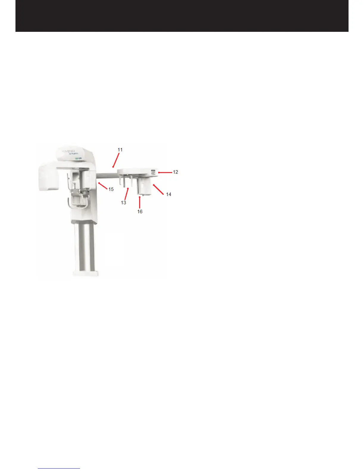
Overview of the medical device: Cephalometric extension:
The Cephalometric version of X-MIND trium medical device consists of the parts listed above and of the following additional
parts:
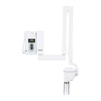
11. CEPH arm extension. Can be positioned both on the right or left side of the vertical column.
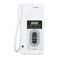
12. CEPH control panel. Provides an intuitive overview of the system to move the mobile column and activate the X-MIND
trium medical device.
13. CEPH patient support. Allows stabilising and immobilising the patient during CEPH exams, by means of ears rest
and nasion rest.
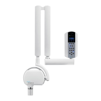
14. CEPH detector sliding group. Enables to slide the detector to follow the X-Ray beam.
15. CEPH secondary collimator. Is positioned on U-arm; it translates during X-Rays (keeping aligned the X-Ray beam
emerging from the tube head with the CEPH detector).
16. CEPH detector. This at panel detector is indicated for use in generating radiographic images of the maxillo-facial region,
more specically it is dedicated for CEPH acquisitions. It gives 2D images of the whole patient head; this detector can
optionally be moved from the CEPH arm to the detector sliding group for PAN examinations.