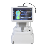
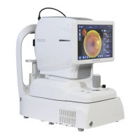
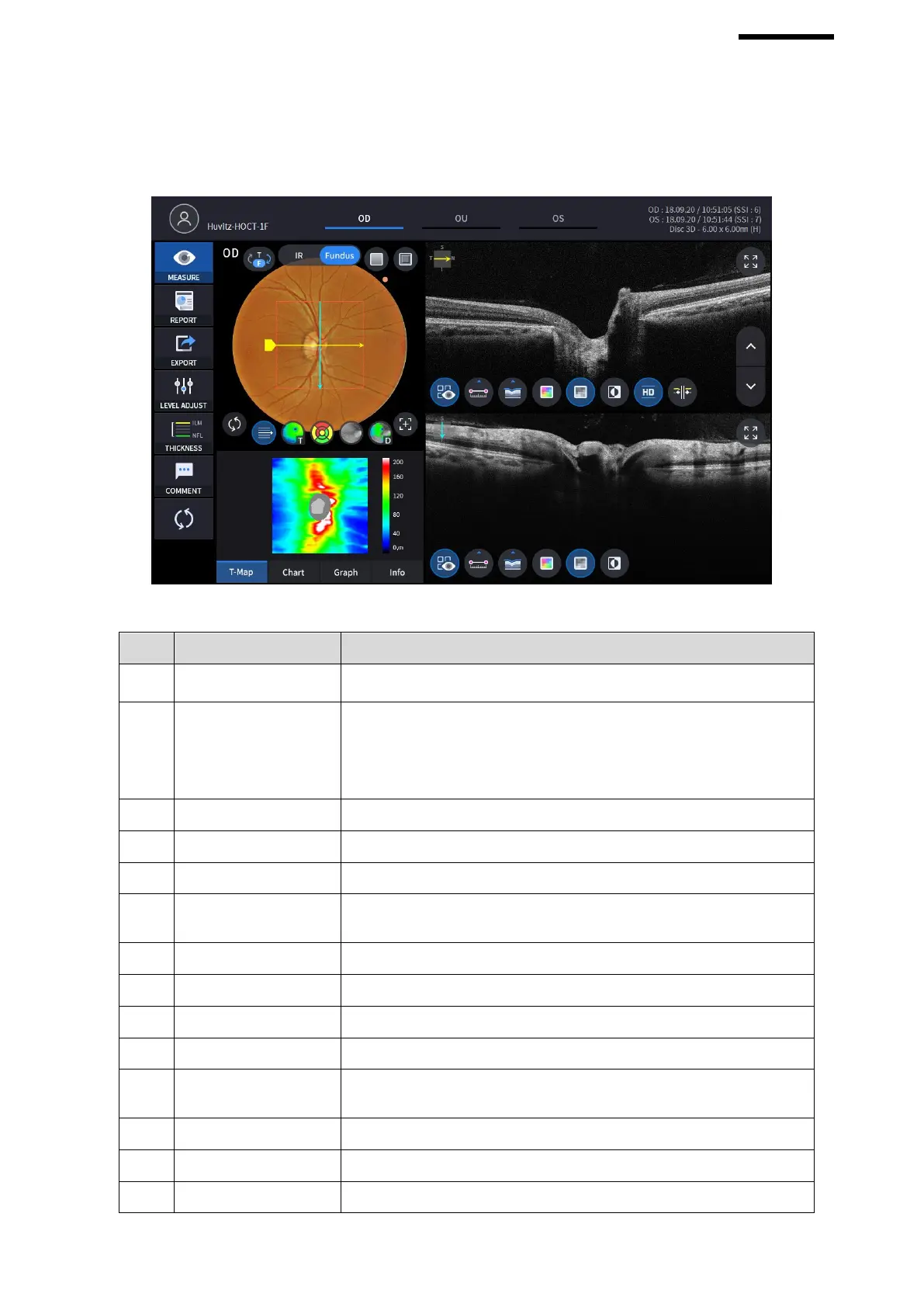
6.7.3. OCT Disc 3D Analysis screen
1. Composition of screen.
Shows the information of patient ID and name. Go back to patient list by
clicking the icon.
Indicates which side of eye is showing.
You can move to the measurement of the other side or the both sides by
selecting unhighlighted tabs.
- OD: right eye, OS: left eye, OU: both eyes.
Displays the date and information that the measurement was taken.
Moves to capture screen after finishing analysis.
Moves to report screen of the current measurement.
If an external storage device is connected, you can store the data that
you want to on an external storage device.
Adjust contrast of Bscan.
Select the analysis range between ILM <-> NFL / ILM <-> RPE.
Leave a brief comment on the patient or measurement.
Select between IR Fundus/Color Fundus if color fundus result is
available.
Apply a red free or embossing effect to the Fundus image.
Moves RNFL Chart center to the center of pattern domain.
Displays Scan direction and position, Enface, Thickness Map,
 Loading...