1
IVIS
®
Spectrum Hardware Manual
1
Welcome
About the IVIS Spectrum Imaging System . . . . . . . . . . . . . . . . 1
IVIS Imaging Systems Technology Support . . . . . . . . . . . . . . . 3
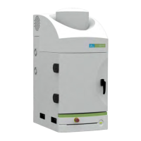
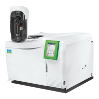
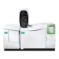
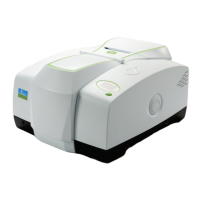
1.1 About the IVIS Spectrum Imaging System
The IVIS
®
Spectrum Imaging System is a high-sensitivity, low noise, in vivo
imaging technology platform that enables noninvasive visualization and
tracking of cellular and genetic activity within a living organism in real time
(Figure 1.1). The system provides both bioluminescence and fluorescence
imaging capability.
For fluorescence imaging, the instrument can operate in reflectance or
transillumination mode. Filtered light from a broad-band lamp provides the
excitation source in both modes. In the reflectance mode, light is delivered to
four reflectors that are located on the ceiling of the imaging chamber. In the
transillumination mode, the excitation light is delivered to an x-y translation
assembly under the stage and focused to a 2 mm diameter beam that can be
directed to a particular location on the underside of the animal subject. The
system includes ten excitation and 18 emission filters that enable spectral
scanning of reporters over the range from 480-850 nm.
3D Tomography The system also includes a structured light projector that enables reconstruction
of the surface topography. The 3D location and concentration of fluorescent
sources can be computed from structured light and transillumination fluorescent
images. The 3D location and strength of luminescent sources is computed from
structured light and luminescent image data.
 Loading...