Operative Technique
Fig. 3
Fig. 5
Fig. 4
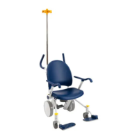
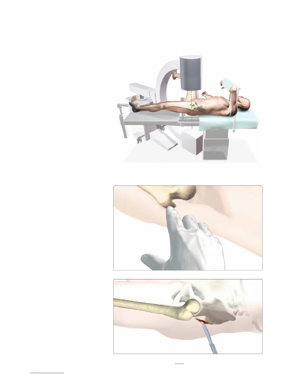
Patient positioning for T2 Recon
Nail insertion is surgeon dependent.
However, it is recommended that
patients are positioned in either the
supine or lateral position on a fracture
table to allow closed reduction of the
fracture (Fig. 3).
Manipulate and reduce the fracture
in the usual fashion, according to the
fracture type. Reduction should be
achieved as anatomically as possible.
If this is not possible, reduction in
one plane should be complete, leaving
reduction in the other plane to be
achieved prior to reaming and nail
insertion.
The unaffected leg is abducted as far
as possible to ease image intensifi er
positioning. This will also allow easier
access to entry point.
Patient Positioning and Fracture Reduction
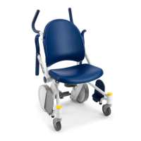
Incision
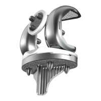
The design of the T2 Recon Nail, with
a 4° medial lateral bend, will only
allow for insertion through the tip of
the greater trochanter.
With experience, the tip of the greater
trochanter can be identifi ed by
palpation (Fig. 4).
A longitudinal skin incision of
approximately 3−5cm is made starting
just above the greater trochanter to the
iliac crest (Fig. 5). The incision is then
deepened to expose the tip of greater
trochanter.
Smaller or larger incisions may be
used based on individual patients
anatomy and at the surgeon’s
discretion.
Note:
The targeting instruments of the
T2ReconNailhavebeendesigned
to allow for a more percutaneous
approach.
11