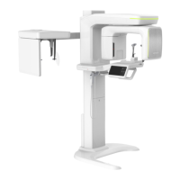
15. Appendices
Green Smart User Manual 181
ENGLISH
Anterior-Posterior Positioning Error
When the arches are positioned incorrectly in the anterior-posterior direction, distortion or
ghosting of the anterior anatomy occurs. Unerupted teeth in the anterior region may not
be imaged on the radiograph if positioned outside of the focal trough. It is important to
note that an error of only 3 mm to 4 mm in either direction will result in a significantly
compromised image.
11
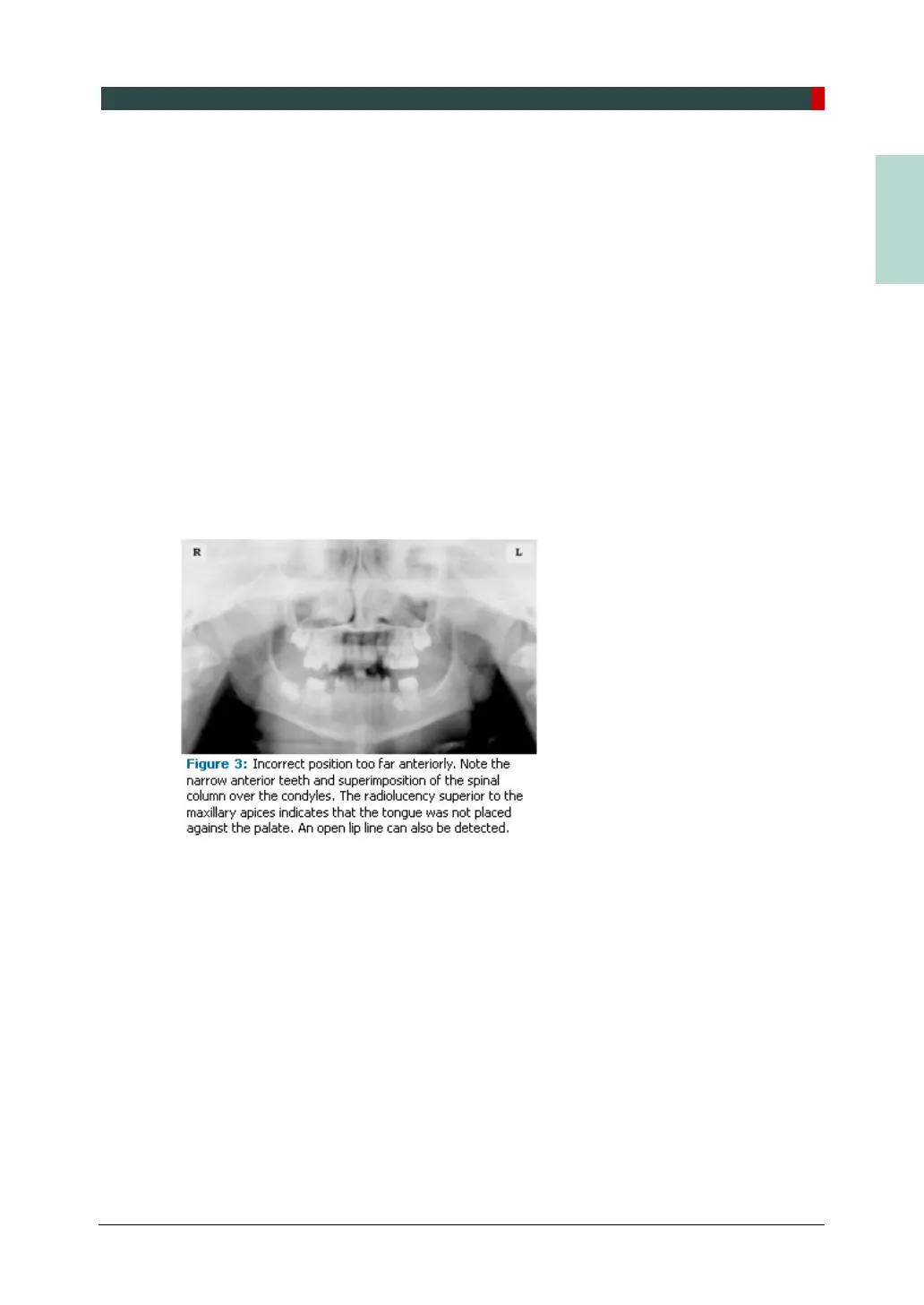
When the arches are positioned too far anterior, the anterior teeth
will appear narrow and diminished in size. The vertebrae of the spinal column may be
superimposed over the condyles at the edges of the film and, depending on the size of the
child, may be superimposed over the rami of the mandible blocking a clear view of the
posterior teeth (Figure 3). When the arches are positioned too far posteriorly, the anterior
teeth will appear broad or widened. If the position is excessively posterior, anterior teeth
may be completely blurred from the image and the condyles may be cut off from the
edges of the film.
To avoid these imaging errors, the anterior teeth must occlude edge-to-edge onto the
designated area of the bite block. Achieving this position is easily compromised during
exfoliation of primary teeth, making precise occlusion difficult when one tooth or multiple
teeth are missing or partially erupted. A cotton roll may be attached to the bite block to fill
in the space created by the missing tooth or teeth. Additionally, an adjustment may be
necessary when using a laser light beam guide. The manufacturer's instructions for
directing the laser light beam at a predetermined tooth or interproximal space usually
apply to adult patients. These instructions may need to be modified for the pediatric
patient with primary or mixed dentition.