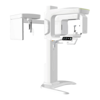
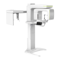
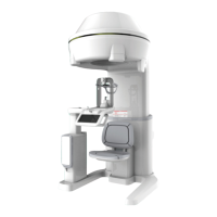
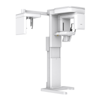
8. Acquiring CEPH Images (Optional)
Green Smart User Manual 83
8. Acquiring CEPH Images (Optional)
8.1 CEPH Imaging Program Overview
Result Images
It provides conventional 2D cephalometric images.
Image Acquisition Method
It acquires multiple images by scanning the specific oral & maxilocial regions with the linear
movement of the narrow detector and reconstructs them to a single 2D image through
computer calculations.
Examination Programs
It is classified as below based on the ROI (Region of Interest).
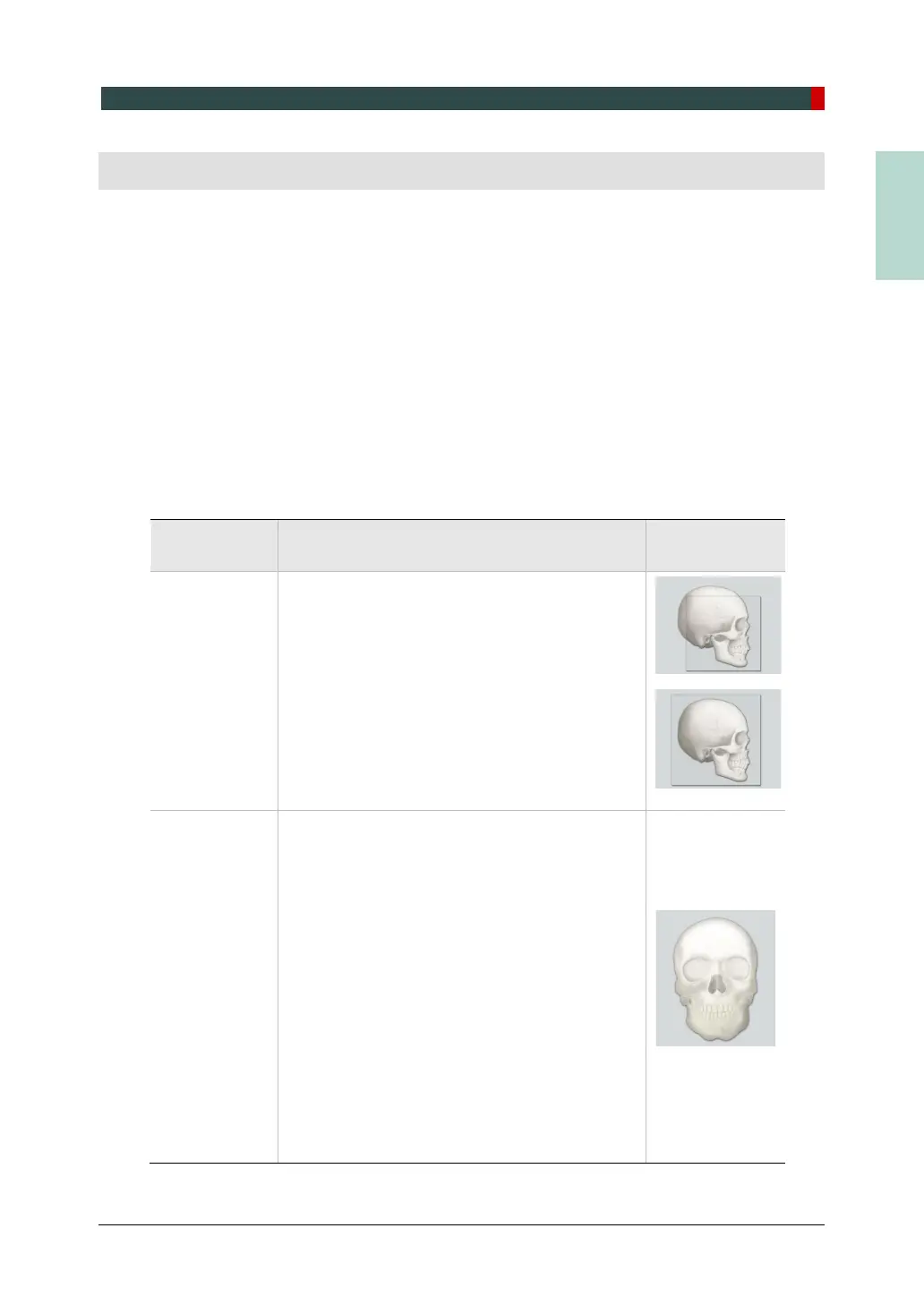
Description Position
Lateral /
Full Lateral
(Optional)
Used to study craniofacial disease, trauma,
and congenital malformation and examine
the soft tissue in the otorhinolaryngological
area, the sinus, and the hard palate.
Measures the angles formed by the
connecting lines between the cranial
measurement points to further assess the
growth of the facial region. It's widely used in
Orthodontics and Oral and Maxillofacial
Surgery.
<Lateral>
PA
The radiation is directed from the posterior of
the skull to the anterior.
Used to examine cranial diseases, trauma,
and congenital malformations.
Used to assess the growth of the lateral side
of the face. It is also used to examine the
ramus mandibulae, the posterior region of
the third largest molar in the lower jaw, the
sidewall of the maxillary sinus, the frontal
sinus, antrum ethmoidale, olfactory pits, and
optic disc pits.
Measures the angles formed by the
connecting lines between the cranial
measurement points to further assess the
growth of the facial region. It is widely used
in Orthodontics and Oral and Maxillofacial
Surgery.
<PA>