15. Appendices
184 Green Smart User Manual
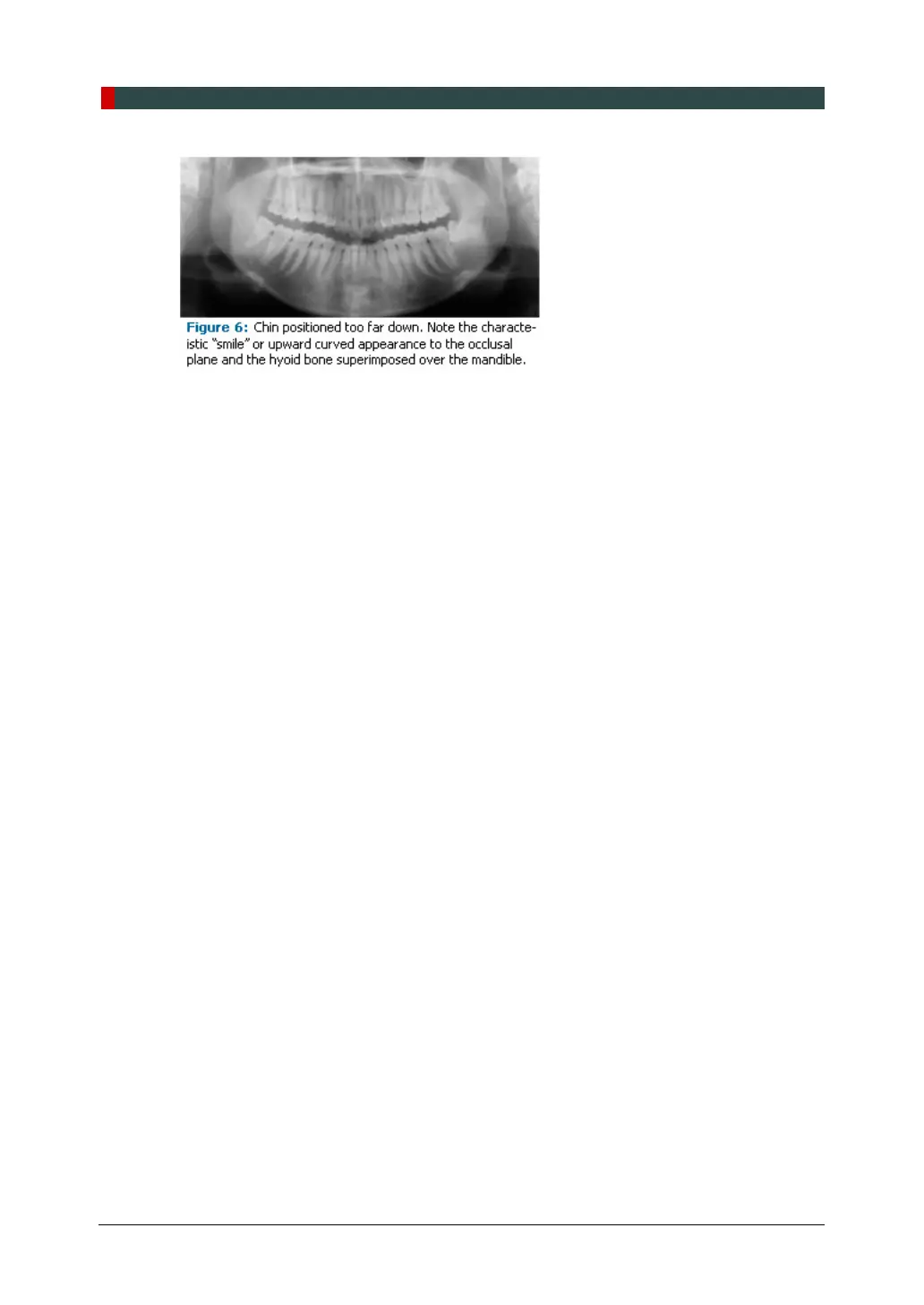
Correct positioning of the arches in the superior-inferior dimension requires that the patient
stands with erect posture while tucking the chin in and down slightly, a direction that both
adults and pediatric patients often find difficult to follow without specific guidance. The
result is often a slumped position with the patient hunching the neck and shoulders over in
an attempt to place the chin on the chin rest. The vertebrae collapse causing attenuation
of the x-ray beam that produces a triangular radiopacity superimposed over the mandible,
and if severe, over the anterior maxillary regions as well.
Depending on the manufacturer, panoramic x-ray machines direct the operator to position
the Frankfort or the canthomeatal plane parallel to the floor, or the ala-tragus line 5° down
toward the floor. This is achieved by raising or lowering the chin rest so that the
appropriate landmark lines up with indicators on the machine (Figure 2). The patient
should be directed to stand in front of the panoramic x-ray machine allowing the operator
to place the chin rest in a position that is slightly higher than the patient's chin. The patient
is then requested to move into the overhead assembly of the machine and remain
standing tall. If further adjustment is needed, it is usually to a lowered chin position. Once
the patient's chin is resting on the chin rest, it is easier to move to a lower position than to
a higher one. To assist with placing the chin on the chin rest while maintaining an erect
posture, the pediatric patient can be directed to stand like a soldier. Most children are
familiar with the straight back, chest forward tucked chin position demonstrated by military
persons, and can readily mimic this stance.
Further Recommendations
Prior to beginning the exposure, the patient should be directed to close the lips around the
bite block and to place the tongue against the palate. Leaving the lips open will create a
soft tissue shadow across the teeth that that can be mistaken for caries.
7
Leaving the
tongue at rest during the exposure allows the radiation to easily penetrate the space of
the oral cavity between the dorsal surface of the tongue and the palate, producing a
radiolucent shadow that diminishes the diagnostic quality of the radiograph (Figure 3).