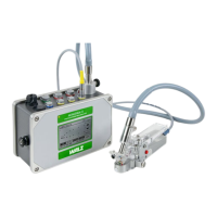
CHAPTER 12 MEASUREMENTS WITH THE MINI-PAM
This leads to strong "energy-dependent" nonphotochemical
fluorescence quenching during the first minutes of illumination
(characterized by low Fm'-values), which partially declines again
when CO
2
-fixation takes over and ATP is consumed.
In order to record an INDUCTION CURVE with the MINI-PAM,
a fixed geometry between sample and fiberoptics must be assured for
the duration of the recording. The recording is started by MODE-
menu function 21: IND.CURVE. It is also possible to record the
light/dark recovery in addition to the dark/light induction
(22:IND.CURVE+REC). In this case information on post-
illumination reactions are obtained, in particular on the recovery of
various components of nonphotochemical quenching (see 12.3.9),
the extent of photoinhibition and also on dark electron flow between
stroma (or cytoplasma) and the electron carrier in the thylakoid
membrane.
Induction curves are either recorded via the analog output of the
MINI-PAM using a chart recorder or via the RS 232 interface using a
PC under WinControl-software. The latter offers the possibility of
online registration and display of various derived fluorescence
parameters, like effective quantum yield and quenching coefficients
(see separate WinControl manual).
Before recording of the actual induction curve, a single saturation
pulse is applied for assessment of Fo, Fm and Fv/Fm after dark
adaptation. This is a prerequisite for correct quenching analysis (see
12.3.1, 12.3.4 , 12.3.6). The delay between this saturation pulse and
onset of illumination can be varied (23: IND.DELAY); its default
value is 40 s. Another variable is the time interval between two
consecutive saturation pulses during actinic illumination (24: IND.-
WIDTH), with a default setting of 20 s.
Due to the outstanding role of molecular O
2
during the induction
period, O
2
partial pressures within the sample has a strong influence
84