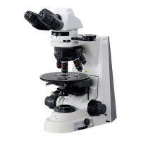
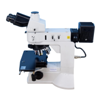
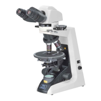
IV Microscopy (Detailed Procedure)
22
z Conoscopic Microscopy
In the case of conoscopic microscopy, the condenser aperture diaphragm functions as a field
diaphragm on the conoscopic image surface. Stop down the diaphragm until it circumscribes
the circumference of the field of view of the conoscopic image (pupil of the objective).
z Orthoscopic Observation/Conoscopic Observation
The following provides an explanation of the characteristic observation method of polarizing
microscopes along with the microscopy procedure. If normal microscopy has not yet been
completed, refer to the previous section and complete normal microscopy.
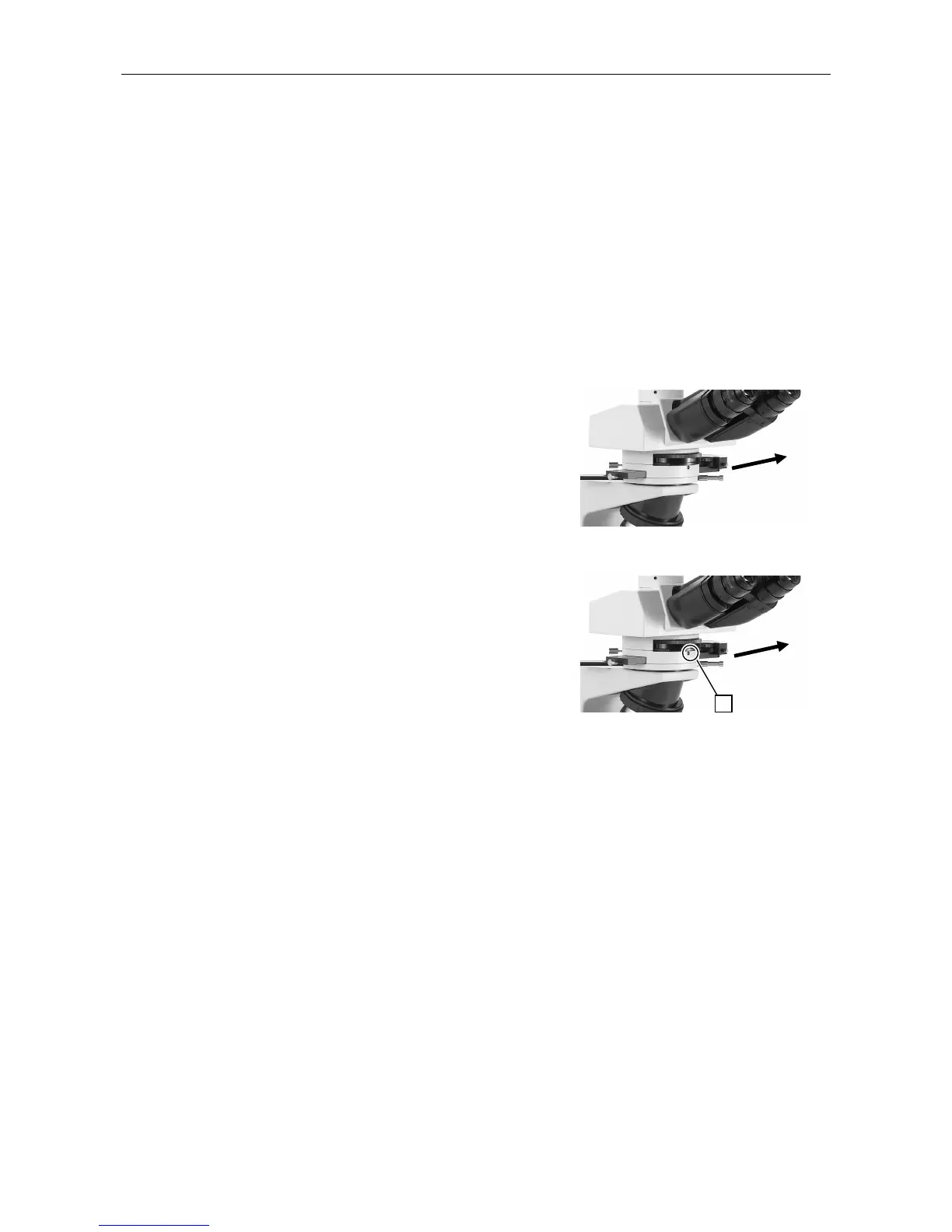
Orthoscopic Observation
In this method, the specimen is observed with
the polarizer and analyzer placed in the optical
path. In this case, the shape of the specimen
is visible (direction of optical axis) and the
optical properties relative to the direction of the
thickness of the specimen can be observed.
• Operation
Pull out the analyzer slider to the right and
move the analyzer into the optical path.
Conoscopic Observation
In this method, in addition to the polarizer and
analyzer, the Bertrand lens is also moved into
the optical path when observing a specimen.
Specimens can be observed from various angles
with diascopic light in the form of a single
image. Differing from orthoscopic observation,
however, the shape of the specimen itself is not
visible with this observation.
• Operation
Pull out the analyzer slider to the right and
move the analyzer into the optical path.
Place the Bertrand lens turret of the
intermediate tube in position “B” to move
the Bertrand lens into the optical path.
(Refer to p. 25 for details regarding the
focusing procedure.) Place the P-CL 1/4λ &
tint plate in the hollow position.
Use an objective having a large numerical
aperture (high magnification: normally 40x
or higher).
Analyzer in the optical path
Analyzer and Bertrand lens
in the optical path
Pull out
Pull out
B