Chapter 2 Microscopy
2.5 In-focus Observation with PFS
50
1
Focus on the specimen with bright-field microscopy or phase contrast microscopy.
Set up for bright-field microscopy or phase contrast
microscopy, as described in Section 2.2,
“Bright-Field (BF) Microscopy,” Section 2.3, “Phase
Contrast (Ph) Microscopy,” or Section 2.4, “External
Phase Contrast Microscopy.”
2
Set up the perfect focus system.
Coarse
Fine
ExFine
Obj.
ON
OFF
6V30W
MAX.
12V100W
L80
EYE
DISPLAY
MEMORY
PFS
ON
RECALL
Z
-
RESET
BRIGHTNESS
R100
L100
FOCUS
B
Coarse
Fine
ExFine
Epi Shutter
FL Block
Refocus
Escape
PFS
OFFSET
OUT
D
ICHROIC
M
I
R
ROR
IN
L80
EYE
MEMORY
DISPLAY
ON
Z
-
RESET
1X
1.5X
BRIGHTNESS
R100
L100
FOCUS
PFS
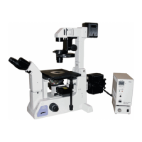
1. Move a PFS objective with the desired
magnification into the optical path by
pressing the Obj. switch on the left operation
panel.
2. Attach an IR filter (infrared filter) to the filter
sliders on the pillar illuminator, and move it
into the optical path.
PFS uses near-infrared light for focus
management. Be sure to place the IR filter into
the optical path to minimize the effect of the heat
rays (infrared light) emitted by the illumination.
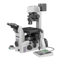
3. Display the PFS information on the status
display panel by pressing the DISPLAY
switch on the front operation panel.
At this point, “PFS: Out” will be displayed.
Display example for PFS information
____________10x/0.25
E100_ Coarse__PFS:Out
4. Move the dichroic mirror into the optical path
by moving the DICHROIC MIRROR - IN/OUT
lever on the top of the PFS Motorized
Nosepiece to the “IN” position.
The PFS information display will change to “PFS:
Off”.
Display example for PFS information
____________10x/0.25
E100_ Coarse__PFS:Off
If “PFS: ER1” is displayed, the objective has not
been configured properly. Check and register the
objective. Refer to the list of supported
objectives on page 127, and to Chapter 4,
“Assembly”.
2-1
2-2
2-3
2-4