EasyManua.ls Logo

Olympus IX70 User Manual

Olympus IX70
31 pages
To Next Page IconTo Next Page
Page #1 background imageLoading...
Page #1 background image
UC BSD Light Microscopy Core Facility Vytas Bindokas, Director
Olympus IX70 Multi-parameter Fluorescence Microscope
Operations Manual (version 1.0 February ‘09)
The IX70 microscope is a manual stand with the ability to collect multi-color fluorescence, differential
interference contrast (DIC) and total internal reflection fluorescence (TIRF), as well as provide optical
sectioning (z-axis stepping) and time lapse imaging. It is equipped with a high sensitivity, high resolution
chilled CCD camera, a green/red or blue/red emission beam splitter for simultaneous two color capture
and MetaMorph + MetaFluor software to control the system.
Examples of types of images that can be captured range from simple image capture (bright field or
fluorescence), sequential capture of up to 4 fluorescent probes, time lapse imaging, (relatively) high speed
imaging, CFP-YFP FRET studies, ratiometric indicator dyes such the calcium probe Fura-2, etc.
Software provides a wide range of image capture and image processing capabilities. MetaFluor is
optimized for real-time (ratio) image capture with real time region of interest (ROI) intensity/
ratio/calibrated data display. MetaMorph is a more general image capture, processing, morphometry
package. Both packages allow images to be automatically collected and annotated.
PDF Created with deskPDF PDF Writer - Trial :: http://www.docudesk.com

Other manuals for Olympus IX70

Question and Answer IconNeed help?

Do you have a question about the Olympus IX70 and is the answer not in the manual?

Olympus IX70 Specifications

General IconGeneral
BrandOlympus
ModelIX70
CategoryMicroscope
LanguageEnglish

Summary

System Operation

Turning on the System

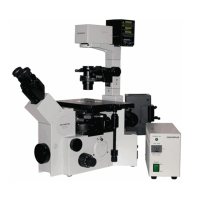
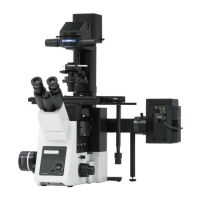
Step-by-step instructions for powering up the microscope and associated equipment.

Shutting down the System

Procedures for safely powering down the microscope system after use.

Differential Interference Contrast (DIC)

DIC Setup and Usage

Instructions for setting up and using DIC optics for contrast in unstained materials.

Fluorescence Imaging

Fluorescence Filter Cubes

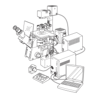
Explanation of filter cube positions and their roles in fluorescence excitation and emission.

MetaMorph Software Usage

Fluorescence Image Capture

Guide to using MetaMorph for capturing fluorescence images, including taskbar functions.

Advanced Imaging Techniques

Total Internal Reflection Fluorescence (TIRF)

Introduction to TIRF microscopy, its principles, and required objectives.

TIRF Operation and Safety

Instructions for initiating TIRF, laser safety, and using the reflection shield.

TIRF Shutdown

Procedure for safely powering down the TIRF laser system and returning components.

TIRF Troubleshooting

Common problems encountered during TIRF imaging and their solutions.

Multi Dimensional Acquisition (MDA)

MetaFluor Software

Troubleshooting

No Image by eye

Steps to resolve issues when no image is visible or illumination is absent.

Ugly Image Issues

Guidance for addressing image quality problems when the system appears functional.

Related product manuals