Important Information — Please Read Before Use
7
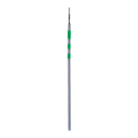
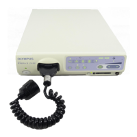
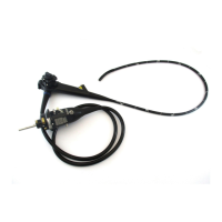
VISERA RHINO-LARYNGO VIDEOSCOPE ENF-V2
Pay special attention to the brightness level setting of the
light source, particularly when operating the electrical
shutter function of a video system. When using a light
source and video system that are compatible with the light
source’s automatic brightness control function, make sure
to use this function. The automatic brightness control
function can keep the illumination light level better.
Refer to the instruction manuals of the light source and
the video system for further details.
Do not continue observation in close proximity to tissue or
keep the distal end of the endoscope in contact with living
tissue for a long time.
When the endoscope will not be used for a long period, be
sure to turn OFF the light source or activate the light
shield function (standby mode, etc.) so that the
endoscope is not illuminated unnecessarily.
• If the endoscopic image becomes dimmer during the
procedure, it may indicate that blood or mucus is adhering to
the light guide on the distal end of the endoscope.
Carefully withdraw the endoscope from the patient and
remove blood or mucus in order to restore optimum
illumination and to ensure the safety of examination. If you
continue to use the endoscope with its obstructed distal end,
the temperature at the distal end may rise and cause
mucosal burns to the patient.
• Never operate the bending section, perform suction, insert or
withdraw the endoscope’s insertion tube without viewing the
endoscopic image. Patient injury can result.
• Be sure to prepare another endoscope to avoid that the
examination must be interrupted due to equipment failure or
malfunction.
• For reasons described below, do not rely on the NBI
1
imaging modality alone for primary detection of lesions or to
make a decision regarding any potential diagnostic or
therapeutic intervention.
It has not been demonstrated to increase the yield or
sensitivity of finding any specific mucosal lesion.
 Loading...