30
Chapter 4 Operation
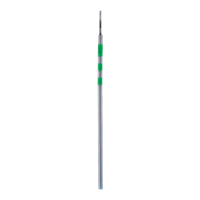
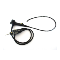
VISERA RHINO-LARYNGO VIDEOSCOPE ENF-V2
Angulation of the distal end
If the angulation control mechanism or any other part of the
system is not functioning properly, stop the procedure
immediately; do not operate the angulation control lever
unless absolutely necessary. Then carefully withdraw the
endoscope while observing the endoscopic image. If the
endoscope cannot be withdrawn from the patient smoothly,
do not attempt to forcibly withdraw it; leave it inside the
patient and immediately contact Olympus. Forcibly
withdrawing the endoscope may cause patient injury.
Operate the UP/DOWN angulation control lever as necessary to guide the distal
end for the insertion and observation.
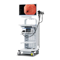
Observation of the endoscopic image
Refer to the light source’s instruction manual for instructions to adjust the
brightness.
Observation of the NBI image
All mucosal areas are to be viewed using traditional white light. NBI
1
imaging
should not be used as a substitute for a thorough traditional examination of the
mucosa.
4.2 Withdrawal of the endoscope
If it becomes impossible to withdraw the transnasally inserted
endoscope, pull its distal end out of the mouth, cut the
flexible tube using wire cutters and, after ensuring that the cut
section will not injure the body cavity or nasal cavity of the
patient, withdraw the endoscope carefully. Therefore, always
prepare wire cutters in advance.
Carefully withdraw the endoscope while observing the endoscopic image.
1 NBI stands for Narrow Band Imaging. For more details, refer to the
instruction manual of the video system center CV-180 or OTV-S7Pro
2
.
2 This product may not be available in some areas.
 Loading...