EN
ONE – User manual Page 5/44
1 Introduction
You have just received your ONE new generation digital intra-oral radiology kit, with direct USB connection. We thank you for
the confidence you have in us and hope that this product will give you entire satisfaction.
We recommend you to read this manual thoroughly before installation; following the guidelines for installation and usage
described in it will exclude risks to the patient and the care team. Please keep it close to your equipment so you can refer to it at
a later date.
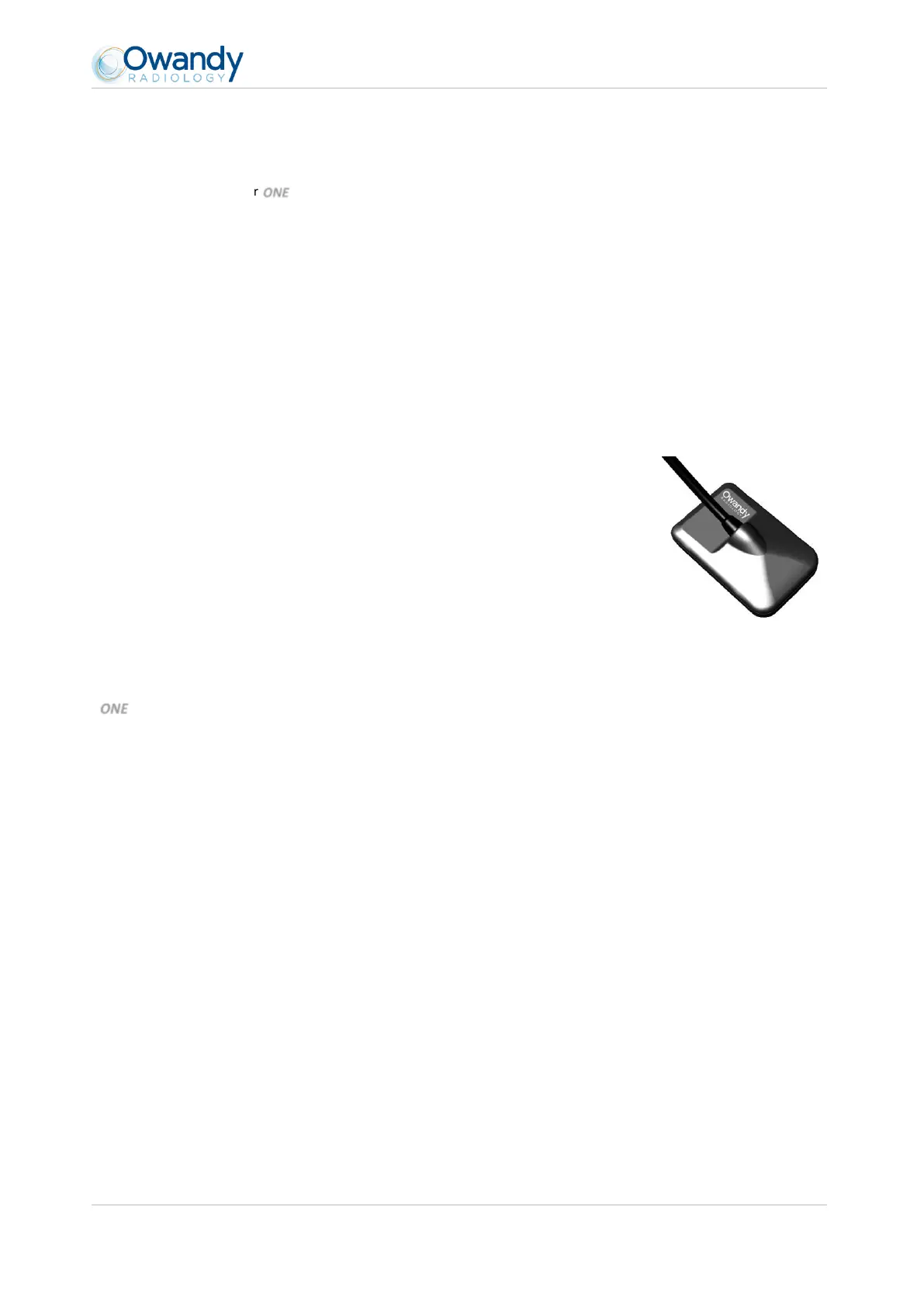
Your sensor uses an X-ray sensitive electronic detector (the flat part at the bottom of the sensor) that replaces the conventional
film used for the acquisition of radiological intra-oral images. The X-rays are automatically detected by the sensor which triggers
image acquisition. The acquired image is displayed almost instantaneously on the screen of the computer to which the sensor is
connected. These digital images can then be manipulated, analysed, saved as files or printed.
The development process of conventional films is thus completely eliminated as well as the possible influences on image quality;
such as the type and age of the chemical product, the temperature of the baths or the development time.
The sensor is available in two sizes; depending on the kit you have ordered you received a size
1, a size 2 sensor or both:
The size 1 sensor allows you to acquire the majority of intra-oral images (peri-apical
and retro-coronary) both vertically and horizontally.
The size 2 sensor furthermore allows you to easily acquire horizontal “bitewing”
images.
The instructions and information in this manual refer to both sensor sizes, unless specifically
stated. The size of the sensor is marked on the sensor itself.
1.1 Intended use
ONE intraoral sensor is an hand held device used to provide digital images of human oral tissues and teeth without
the use of a conventional x-ray film.
Placed in the oral cavity of the patient, the sensor is subjected to X radiation from an x-ray generator (not part of
the device) a few tenths of a second. The sensor, upon radiation exposure, captures the image that is then
transferred and viewed on the practitioner's computer.
ONE is used for diagnosis purpose by dental practitioners or radiologists.
1.2 Intended patient population
One sensor can be used with the following type of patient:
Age peadiatric to geriatric
Patient status/Health: the patient is conscious
Nationality: multiple
Note: PATIENT is not an OPERATOR