Planmeca Intra X-ray Unit 21
PREMOLAR AND CANINE EXPOSURE
User’s Manual
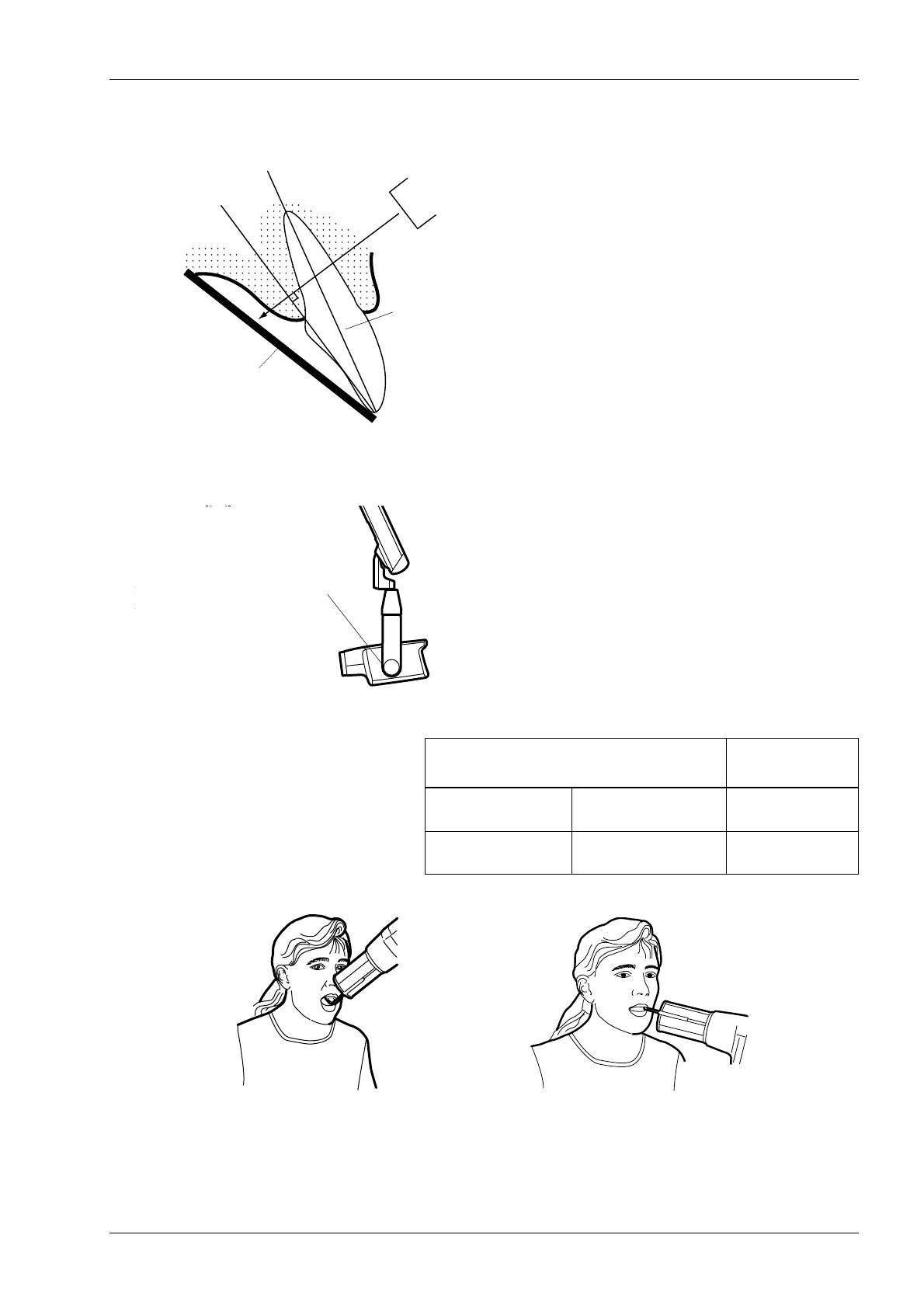
Bisecting angle technique (optional)
The patient holds the film or sensor in place with his
finger. The X-ray beam is directed perpendicularly
towards an imaginary line which bisects the angle
between the film plane and the long axis of the tooth.
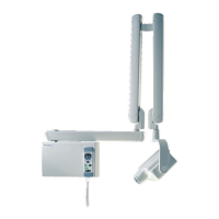
Positioning the cone
The angle of the cone is indicated on the scale located on
the vertical joint of the tube head.
The optional long cone can be attached into the short
cone. Refer to chapter 5.2 “Selecting the cone” on page 8.
Select the cone angle from the table below.
Position the cone according to the figure below.
TEETH
ANGLE OF
INCLINATION
Premolars and
canine teeth
Maxilla +45°
Premolars and
canine teeth
Mandible -10°
Film or
Long axis
of the tooth
sensor
Scale for the cone angle
Mandibular premolar and canine
Maxillary premolar and canine