GMK Sphere Calipered Kinematic Alignment Surgical Technique
6
3. DISTAL FEMORAL RESECTION
Calipered kinematic alignment sets the femoral component
coincident to the distal articular surface of the native femur.
Restoring the native distal femoral line requires
compensations of ~2 mm for worn cartilage when present
on the distal femoral condyles. When measuring the cut,
~1 mm of saw blade kerf should be accounted for.
Compensation for bone wear is rarely required at the 0°
of flexion position on the osteoarthritic femoral condyle
with end-stage varus or valgus deformity.
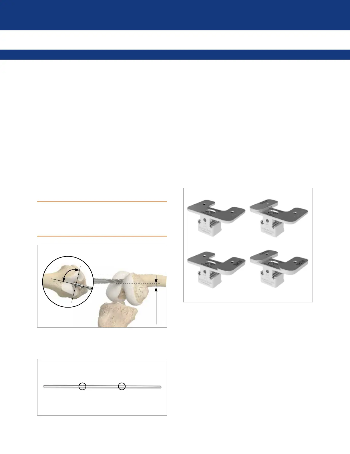
3.1 PLACING THE INTRAMEDULLARY ROD
First, set the flexion-extension position of the femoral
component. Drill a hole midway between the top of the
intercondylar notch and the anterior cortex, depending on
the anterior-posterior size of the femur. Keep a 5-10 mm
bridge of bone between the posterior rim of the drill hole
and the top of the intercondylar notch. Orient the drill
parallel to the anterior surface and perpendicular to the
distal articular surface of the distal femur.
CAUTION
Excessive flexion of the femoral component could lead to
patellar instability. Orienting the drill hole parallel to the
anterior surface of the distal femoral shaft minimizes
flexion of the femoral component.
5-10 mm
4.
Insert the rod 8-10 cm into the femur using the marks
engraved on the shaft as a reference.
5.
Determine the extent of cartilage wear on each distal
femoral condyle. Use a ring curette to remove any partially
worn cartilage on the bone.
Set the varus-valgus angle and proximal-distal level of the
femoral component. This is done using a distal cut
referencing guide. The guide can compensate for 2 mm of
cartilage wear on the worn condyle(s).
4 distal referencing guides are available:
•
1x UNWORN/UNWORN: This is for cases with no
cartilage wear on either distal femoral condyle
•
1x UNWORN/WORN: This is for cases with cartilage
wear on the right medial or left lateral condyle
•
1x WORN/UNWORN: This is for cases with cartilage
wear on the left medial or right lateral condyle
•
1x WORN/WORN: This is for cases with wear on both
distal femoral condyles
6.
Select the appropriate guide depending on the operative
side and the pattern of cartilage wear.
Place it onto the intramedullary rod and advance it until it
contacts both femoral condyles. Stabilize the distal cut
reference guide by means of two threaded pins into the
distal holes.