IV Microscopy (Detailed Procedure)
24
11
Orientation of Polarizing Plates (Polarizer and Analyzer)
(1) Push in the analyzer slider to the right and
move the analyzer out of the optical path.
(2) Focus on the specimen.
(3) Pull out the analyzer knob and move the
analyzer into the optical path.
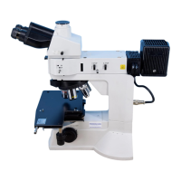
(4) Turn the analyzer rotation ring and align at
the "0" position on the analyzer scale.
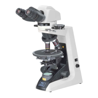
(5) Insert the polarizer beneath the condenser.
(6) Move the specimen out of the optical path.
(7) Move the Bertrand lens into the optical path.
The pupil of the objective will then be visible
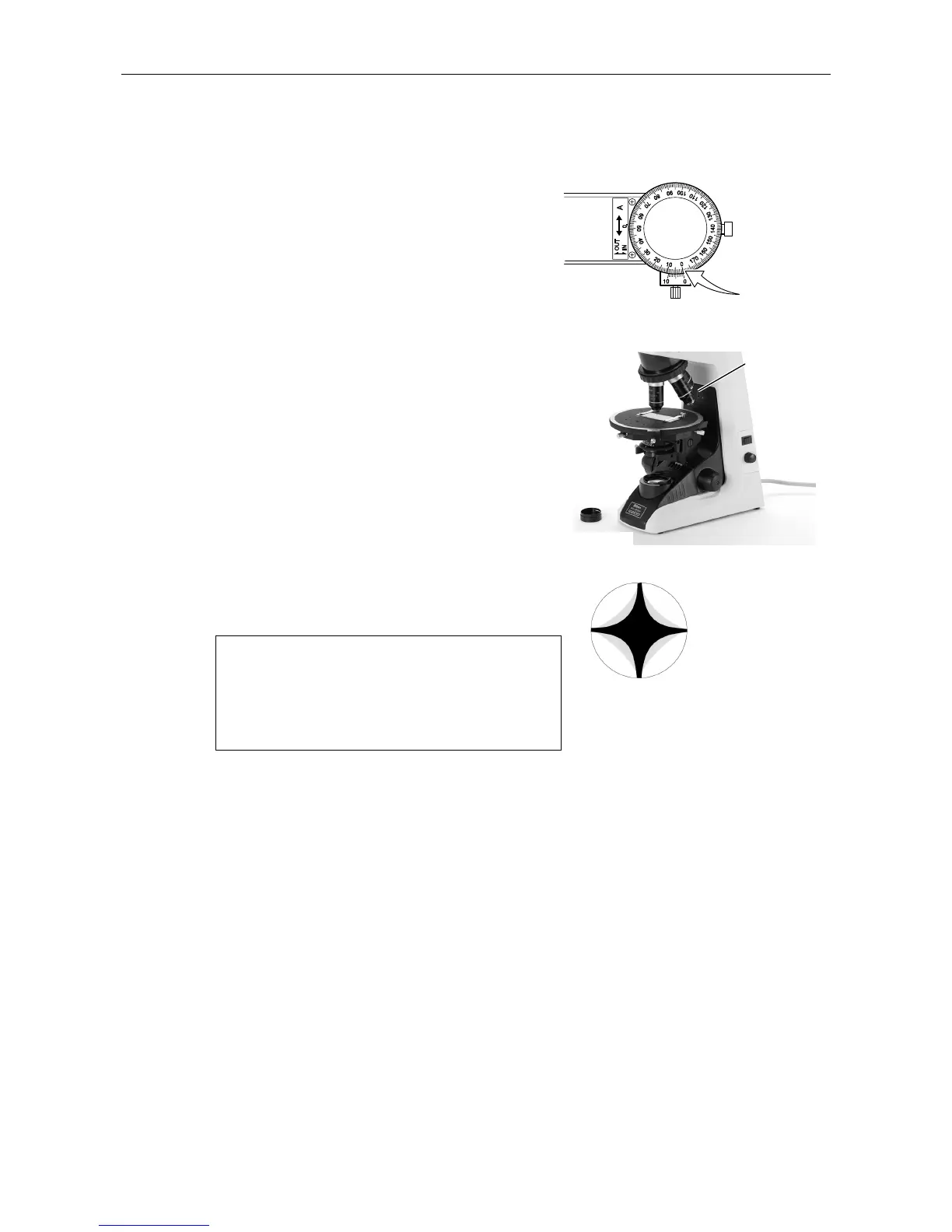
through the eyepiece. Turn the polarizer and
adjust so that the dark cross image appears
in the pupil as shown in the figure in the
conoscopic observation. This is so-called
crossed Nicols position, where the directions
of the polarizer and analyzer coincide with
those of the orientation plate on the pillar of
the microscope base (the polarizer is in the X
direction: P and the analyzer is in the Y
direction: A).
It should be noted that the X direction
may be explained as that of the
analyzer and Y direction as that of the
polarizer in some commercially
available technical manuals and
reference books.
Analyzer
scale: 0
Orientation
plate
Polarizer
Dark cross ima