Acquiring a Good Image
KODAK RVG Digital Radiography Systems User’s Guide (CS4000_en) 2–15
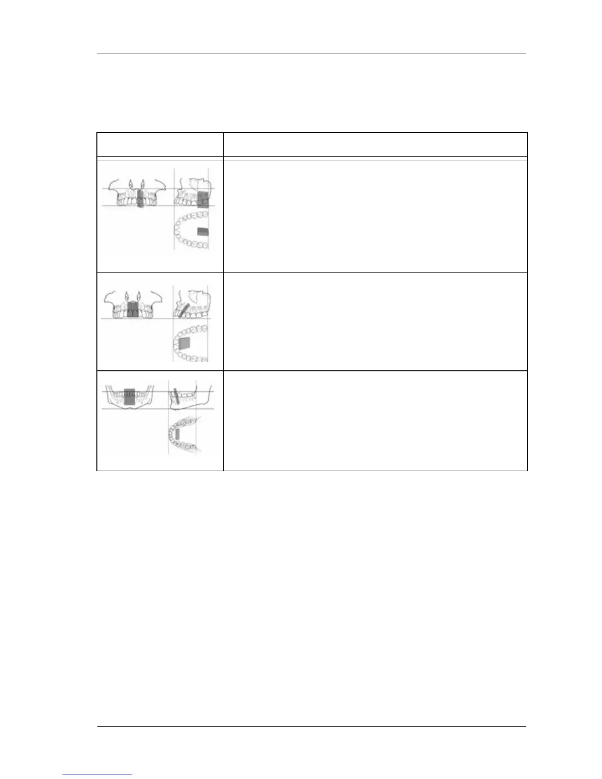
Table 2–2 describes examples of positioning.
Table 2–2 Positioning Examples
Example Description
Upper posterior region
Use the roundness of the palate to place the sensor to frame the
apical area. Use Rinn type positioners for paralleling technique.
Maxillary Anterior region
Use a bisecting technique. Have the patient hold the sensor
against the tooth with a finger. For the paralleling technique,
move the lower part of the sensor away from the incisive edge
to place it parallel to the real axis of the teeth.
Lower Anterior Region
For a narrow mouth, move the sensor back parallel to the real
axis of the teeth while pushing back the tongue slightly. Use the
blunt edges of the sensor to depress the floor of the mouth to
better frame the apical area.