41
Only when no graticule is inserted in both
eyepieces:
• Largely defocus the object or remove it from
the beam path.
• Adjust the eyelens so as to focus on the
boundary of the field of view. When adjusting
the eyelens you will see a bright line around
the body of the eyepiece. This line shows the
correct position of the eye-lens for people
with normal eyesight and for wearers of
glasses who look through the microscope
with corrective glasses.
Remove glasses with bifocal or progressive
lenses before looking through the micro-scope.
• Focus the object through the eyepieces.
Only if one eyepiece has no adjustable
eyelens:
• Focus the object through this eyepiece first
(close your other eye).
• Focus the image again by adjusting the
eyelens of the second eyepiece.
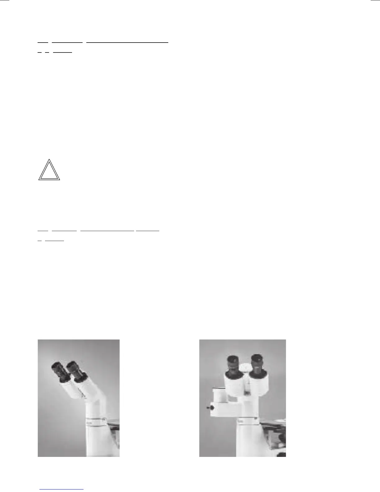
Fig. 45 DMILT tubeFig. 44 DMILB tube
Correction for defective vision
• Look through the right eyepiece with your
right eye and use the fine adjustment to focus
on the specimen.
• Then look onto the same spot of the specimen
with your left eye and rotate the left eyepiece
connection piece until the point on the object
is sharply focused. Do not use the fine
adjustment to achieve this.
• If eyepieces with adjustable eyelenses are
used, do not correct for defective vision by
adjusting an eyepiece tube but by adjusting
the eyelens of the eyepiece.
DM ILT trinocular tube
• Switch the beam splitter to visual obser-vation
by moving the rod. The meaning of the
switching positions is marked with symbols on
the lateral face of the tube.
Rod retracted = visual
Rod inserted = photo
• Adjustment of the eyepieces is made in the
same manner as for the binocular tube.
• Correct for defective vision by adjusting the
eyelens of the eyepiece.


 To Next Page