© Midmark Corporation 2016
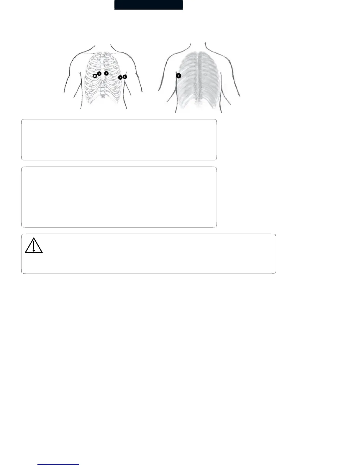
V7 (Blue) – Horizontal level of V4, at the posterior axillary line.
Modified 12-Lead ECG Hookup
Precordial Posterior
The live ECG acquisition screen will show the signal tracings after
all limb leads have been connected. When the right-leg (RL) lead
becomes detached, the system behaves as if all electrodes were
disconnected.
Lead placement does affect the ECG waveform. When the limb
leads are placed on the torso, waveform changes might be seen
in the QRS amplitude, axis shift occurs, Q waves can be seen,
and T waves might appear flipped or flattened. These changes
are clinically significant in that they are associated with cardiac
ischemia. If a non-standard lead placement is used, note the
variation in the ECG comment field.
Caution
Do not use a modified 12-lead ECG placement when performing a STAT ECG. The
automated ECG analysis algorithm assumes the standard 12-lead ECG placement. Any
deviation from the standard 12-lead ECG placements may affect the accuracy of the
automated interpretation.