Image Optimization 5-9
Middle Line:
“Middle Line” is used to locate the focus point of lithotrity wave during lithotrity
treatment. For details, please refer to 12.3 Middle Line
Click [Middle Line] in the image menu at the left side of the screen to turn on the
lithotrity.
LGC
Adjust the gain of scan lines to increase the image lateral resolution.
Click [LGC] to access the adjusting dialog box.
The 8 LGC items displayed on the screen indicate the corresponding image
areas on the image screen.
Click corresponding [LGC1-8] to adjust the gain. The higher the value, the
higher the gain.
The system also provides several preset parameters for imaging.
5.5 M Mode
5.5.1 M Mode Exam Protocol
1. Select a high-quality image during B mode scanning, and adjust to place the area of
interest in the center of the B mode image.
2. Press <M> on the control panel, and roll the trackball to adjust the sampling line.
3. Press <M> on the control panel again or <Update> to enter M mode, then you can
observe the tissue motion along with anatomical images of B mode.
4. During the scanning process, you can also adjust the sampling line accordingly when
necessary.
5. Adjust the image parameters to obtain optimized images.
6. Perform other operations (e.g. measurement and calculation) if necessary.
5.5.2 M Mode Parameters
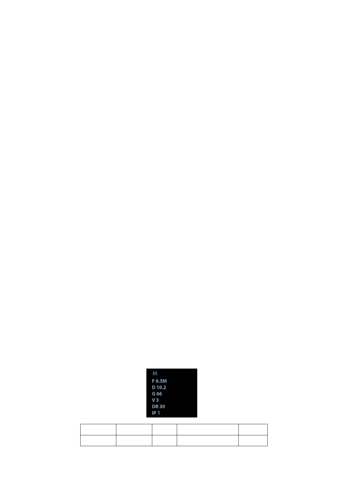
In M mode scanning, the image parameter area in the upper left corner of the screen
displays the real-time parameter values as follows:

 Loading...