ENGLISH
PSPIX² • User manual
6
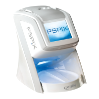
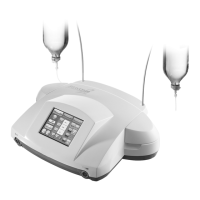
2.3 PRINCIPLE OF OPERATION
The PSPIX system is only intended for the optical reading and processing of IP images. The PSPIX system
converts the image data stored on the IP into a digital image. This image is then transfered by an acquisition
module and displayed on the screen by dental imaging software, and is fi nally viewed and interpreted by
the practitioner.
Stage 1: Acquiring a X-ray image
• The IP is slid into a single-use cover integrated in a hygiene bag, which is also single-use.
• The protected IP is now placed in the patient’s mouth behind the part of the mouth to be X-rayed.
• After exposure, the protected IP is removed from the patient’s mouth and then disinfected with a
disinfectant cloth. The user can then remove his/her gloves and clean his/her hands.
• The cardboard protective cover along with the IP contained inside are taken out of the plastic hygiene
bag by the user.
Stage 2: Reading the image data
• Once the PSPIX system is ready to use, the IP (with it’s protective cover still attached) must be correctly
inserted into the IP slot (1) of the PSPIX system.
• The IP is then detected by the PSPIX system and is automatically logged by the system to be read.
• The protective cover remains in place in the IP slot (1). Once empty, it can be removed and discarded.
• The IP then arrives in the PSPIX system reading module where the images are read with the aid of a laser.
The IP’s are then collected, processed and transferred to the connected computer via an acquisition module.
An animation indicates the progress of the IP reading.
• After several seconds the digital image will appear simultaneously on the Touch screen of the PSPIX (2) and
on the computer screen through the dental imaging software.