6H4130 – September 2007 23
Extraoral Gain Verification
[1] In the Acquisition window, set the button to Extraoral.
[2] Select High Speed scanning mode.
[3] Type the x-ray setting used in your practice for adult patients. See Table 1 for the factory setting for tuning a unit
in production.
[4] Load a freshly erased plate into the cassette.
[5] Do one of the following:
• If using cephalometric imaging mode, attach 6mm-thick AL phantom on the center of the cassette and insert
it into the cassette slot on the ceph x-ray machine.
Note
Using masking tape to secure the phantom.
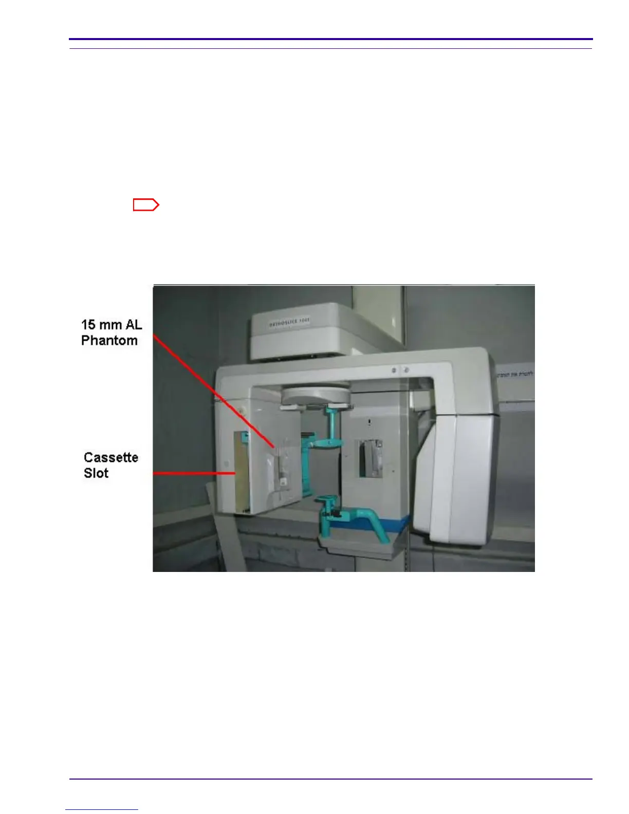
If using panoramic imaging mode, attache 15mm-thick AL phantom on the secondary collimator slot of
the panoramic x-ray machine and the panoramic cassette into the cassette slot as detailed in the
following diagram.
[6] Make an x-ray exposure, and wait 2 minutes after exposing the plate.
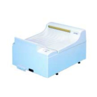
[7] Load the plate into the CR7400 drum and click Scan. As the scan is completed, the image is displayed in the
image diagnostic viewer.
[8] Verify that No-Correction is selected in the Data correction method field.
[9] Do one of the following:
• If using cephalometric imaging mode, measure the pixel value in the center of the image.
• If using panoramic mode, measure the pixel value at the end quarters.
 Loading...