5-20 Image Optimization
5.6 PW/CW Doppler Mode
PW (Pulsed Wave Doppler) mode or CW (Continuous Wave Doppler) mode is used to
provide blood flow velocity and direction utilizing a real-time spectrum display. The
horizontal axis represents time, while the vertical axis represents Doppler frequency shift.
PW mode provides a function for examining flow at one specific site for its velocity,
direction and features. CW mode proves to be much more sensitive to high-velocity flow
display. Thus, a combination of both modes will contribute to a much more accurate
analysis.
Adjustment items, such as SV, Steer, Duplex, Triplex, iTouch and HPRF, are not available
in CW mode.
5.6.1 Basic Procedures for PW/CW Mode Exam
1. Select a high-quality image during B mode or B + Color (Power) mode scanning, and
adjust to position the area of interest in the center of the image.
2. Press <PW>/<CW> to adjust the sampling line,
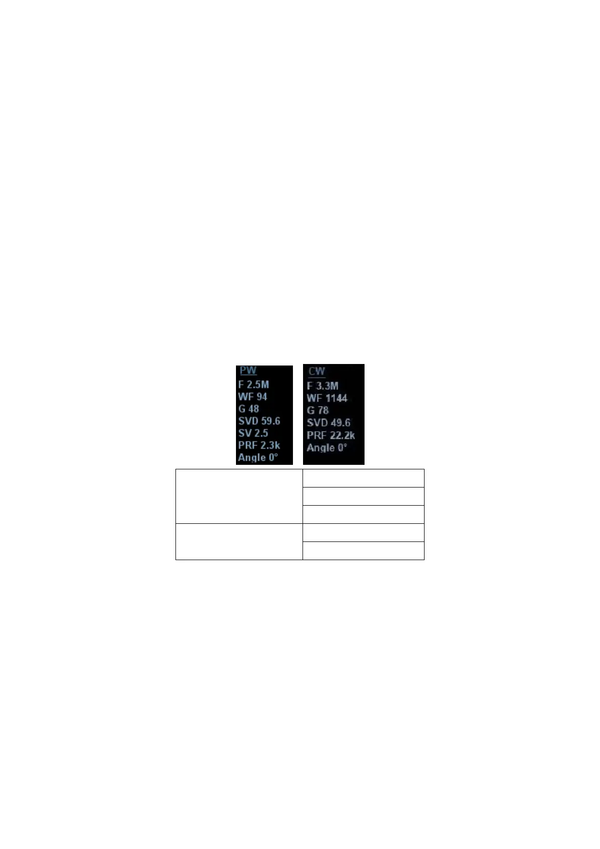
z The sampling status will be displayed in the image parameter area in the top-
right corner of the screen as follows:
PW Sampling Line
Adjustment
SV
Angle
SVD
CW Sampling Line
Adjustment
Angle
CW Focus Depth
3. Set the position of the sample line by moving the trackball left and right. Set the SVD
by moving the trackball up and down. Adjust the angle and SV size according to the
actual situation.
4. Press <PW>/<CW> or <Update> again to enter PW/CW mode and perform the
examination. You can also adjust the SV size, angle and depth in real-time scanning.
5. Adjust the image parameters during PW/CW mode scanning to obtain optimized
images.
6. Perform other operations (e.g., measurement and calculation) if necessary.
If “sampling line displaying” is selected, then the screen will display the sampling line all
the time and pressing <M> one will lead to entering M mode directly. For details, see
“5.1.3 Quickly Saving Image Settings” chapter.