EasyManuals Logo

Mindray Resona 7 Operator's Manual

Mindray Resona 7
492 pages
To Next Page IconTo Next Page
To Previous Page IconTo Previous Page
Page #182 background imageLoading...
Page #182 background image
6 - 56 Operator’s Manual
6 Image Acquisition
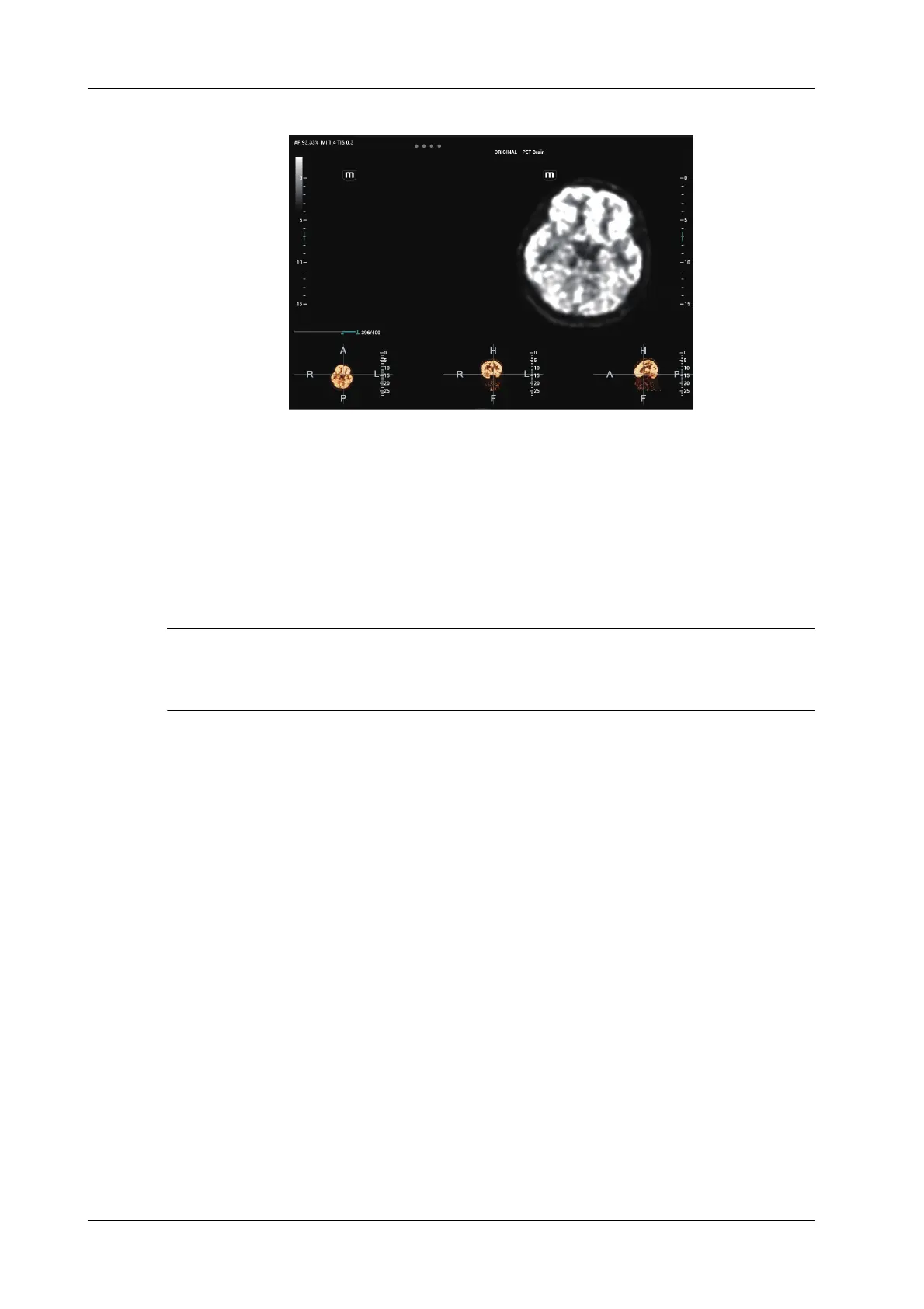
Volume data is loaded to Fusion Imaging exam. The page is given below:
Acquiring Freehand Volume Data
Except importing existing volume data, the operator can also acquire freehand 3D cine on fusion
imaging mode.
6.15.6 Marks
Mark tumor position, lesion position on CT/MR/PET/Freehand image. Be sure of the lesion
appearing on US and CT/MR/PET/Freehand image at the same time after the registration is
completed.
TIP:
Mark the tumor or lesion on CT/MR/PET/Freehand image after CT/MR/PET/Freehand data is
loaded. Generally, it is available to mark the tumor or lesion before/during/after the registration.
Perform the following procedure:
1. Tap [Mark on Volume Data] to enter the page. Select [Add Marks]>[Display Marks] to activate
the mark.

Table of Contents

Other manuals for Mindray Resona 7

Questions and Answers:

Question and Answer IconNeed help?

Do you have a question about the Mindray Resona 7 and is the answer not in the manual?

Mindray Resona 7 Specifications

General IconGeneral
TypeUltrasound System
Power SupplyAC 100-240V, 50/60Hz
Transducer Frequency Range1.0 MHz
Transducer Ports4
Transducer TypesConvex, Linear, Phased Array, Volume
Imaging ModesB-Mode, M-Mode, Color Doppler, Power Doppler, Pulsed Wave Doppler, Continuous Wave Doppler, Tissue Doppler
Advanced Imaging TechnologiesSpeckle Reduction Imaging
ConnectivityDICOM, Ethernet, USB, HDMI
ApplicationsGeneral Imaging, Radiology, Cardiology, Vascular, OB/GYN, Urology, Small Parts, Musculoskeletal
Battery LifeNo battery

Related product manuals