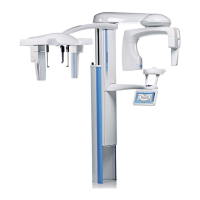
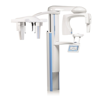
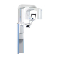
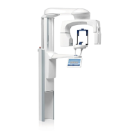
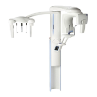
ProMax X-ray unit with Dimax3 1
INTRODUCTION
User’s Manual
1 INTRODUCTION
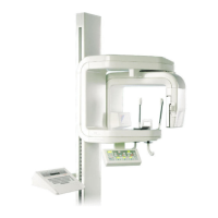
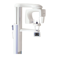
The Planmeca ProMax X-ray unit uses panoramic, linear
tomographic and cephalometric techniques to produce X-
ray images for the diagnosis of dentomaxillofacial
anatomy. The unit is allowed to be used only under
supervision of a dental/health care professional.
This manual describes how to operate the Planmeca
ProMax X-ray unit equipped with the Planmeca Dimax3
digital sensor head. Please read these instructions
thoroughly before using the unit.
NOTE You need a PC with the Dimaxis imaging software
program in order to save, view and modify the
radiographs. The Dimaxis software program has
a separate manual that should be used in
conjunction with this manual.
NOTE The optional ProMax Cephalostat has a separate
manual that should be used in conjunction with
this manual.
The ProMax X-ray unit fulfills the requirements of
Directive 93/42/EEC.
The ProMax X-ray unit fulfills the requirements of
standard EN 55011, class A.
CAUTION Federal law restricts this device to sale by or on
the order of a dentist.
NOTE The unit’s software revision is shown briefly on
the control panel when the unit is switched on.
This manual is valid for software revision 1.19.5
or later. This software revision is compatible with
Dimaxis software revision 3.3.1 or later.
The display values shown in this guide are only examples
and should not be interpreted as recommended values
unless otherwise stated.
The exposure values required to produce good X-ray
images will vary considerably according to the build and
age of the patient. Therefore, the exposure values given
in this guide are average values and are only meant to
guide the user. Users are encouraged to develop their
own radiographic techniques based on these values.
The kV value can be reduced by 2 kV from the suggested
value to improve the image contrast. The radiation dose
(mA) can be reduced by 20-40% without significantly
lowering the image quality.
Make sure that you are fully acquainted with the
appropriate radiation protection measures and these
instructions before using the unit.
 Loading...