How to display X-ray images
The HI-CAT color- and HI-CAT black and white images
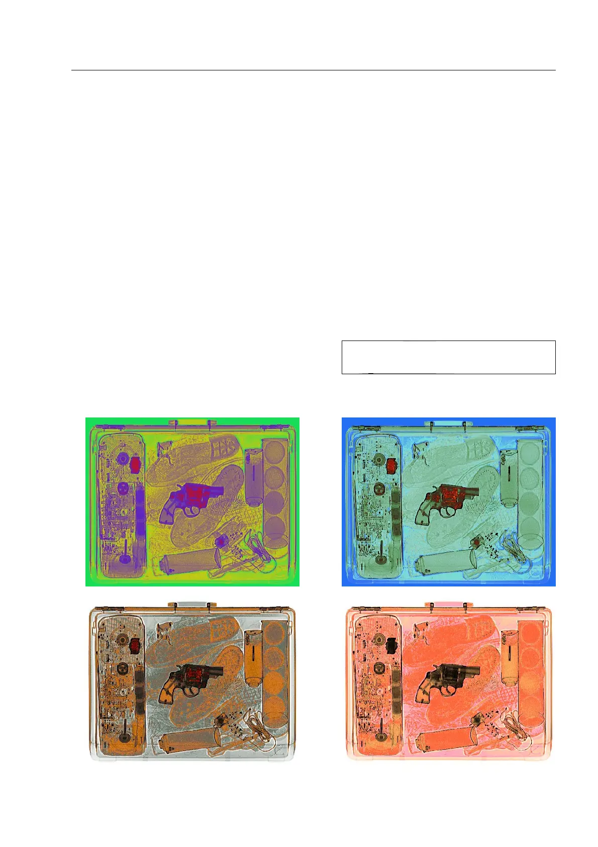
Use the HI-CAT system to generate 6 different color images (HI-CAT 1...6) and 2 different black and white im-
ages (HI-CAT 7 and 8).
HI-CAT color and black and white images can only be selected via priority keys assigned to the functions
HI-CAT1...8, or they must be defined as default image display.
The color images
In the various HI-CAT color images the varying absorption degrees of the scanned object are displayed in
greatly different colors. All color images have one thing in common: there is no direct relation between ab-
sorption and grey levels/brightness and between color and material kind as it is the case in the HI-MAT
PLUS
color image*.
In this case, the contrast of the X-ray image surpasses the
normal B/W and HI-MAT
PLUS
image displays since the hu-
man eye can much better distinguish different colors than
brightness levels which are proportional to the absorption. Object structures will become much better visible
that way.
HI-CAT color image 1 HI-CAT color image 2
HI-CAT color image 3 HI-CAT color image 4
95587412 01/01/09 © Smiths Heimann
1-69
I
The image display is improved by a real-
time sharpening filter.

 Loading...


