Chapter 1: Hyperion Imaging System Introduction and Specifications
Introduction
Hyperion Imaging System User Guide
7
34BHyperion Imaging System Technology
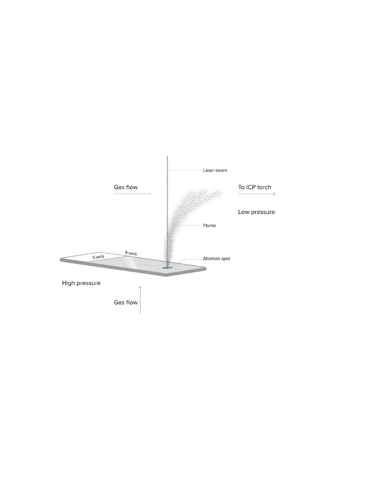
The Hyperion Imaging System technology is an innovative system based on laser ablation
technology coupled with time-of-flight (TOF) mass cytometry of the resulting plume (see
Figures 2 and 3). The system uses a precisely directed laser beam focused at 1 µm to collect
protein markers stained with metal-tagged Maxpar® antibodies and directs these metal tags
for detection using CyTOF® technology. The sample on the slide is ablated and aerosolized.
The resulting aerosol plume is delivered through the coupling tube to the Helios™
inductively coupled plasma (ICP) torch through the argon and helium flow.
Figure 2. A glass slide containing a biological sample is loaded into the Hyperion Imaging System. The beam from
the solid-state laser is directed to the sample on the slide. The resulting plume is directed through the coupling
tube in a stream of argon gas towards the ICP Torch for CyTOF detection.