5-18 Image Optimization
Operations
Click the [Dynamic Range] item on the image menu to adjust the dynamic
range.
Increasing dynamic range will lead to higher sensitivity to low-power signals,
thus enhances the range of signals to display.
5.8 PW Doppler Mode
PW (Pulsed Wave Doppler) mode is used to provide blood flow velocity and direction utilizing
a real-time spectral display. The horizontal axis represents time, while the vertical axis
represents Doppler frequency shift.
PW mode provides a function to examine flow at one specific site for its velocity, direction and
features.
PW is optional.
5.8.1 Basic Procedures for PW Mode Exam
1. Select a high-quality image during B mode scanning.
2. Press the user-defined key for the PW mode to adjust the sampling line,
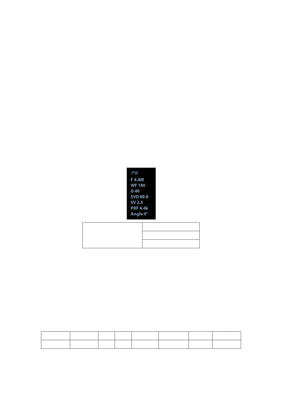
The sampling status will be displayed in the image parameter area in the upper right
corner of the screen as follows:
PW Sampling Line
Adjustment
3. Set the position of the sample line by moving the trackball left and right, and set the SVD
by moving the trackball up and down, adjust the angle and SV size according to the
actual situation.
4. Press the user-defined key for the PW mode or <Update> to enter PW mode again and
perform the examination. You can also adjust the SV size, angle and depth in real-time
scanning.
5. Adjust the image parameters during PW mode scanning to obtain optimized image.
6. Perform other operations (e.g. measurement and calculation) if necessary.
5.8.2 PW Mode Image Parameters
In PW mode scanning, the image parameter area in the upper right corner of the screen
displays the real-time parameter values as follows: