TEMPOROMANDIBULAR JOINT EXPOSURE
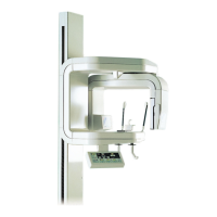
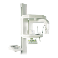
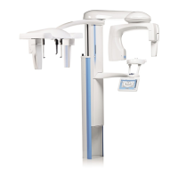
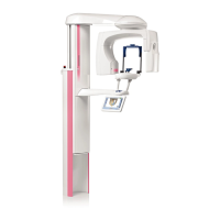
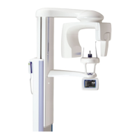
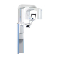
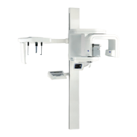
38 Planmeca Proline XC Panoramic X-ray
User's manual
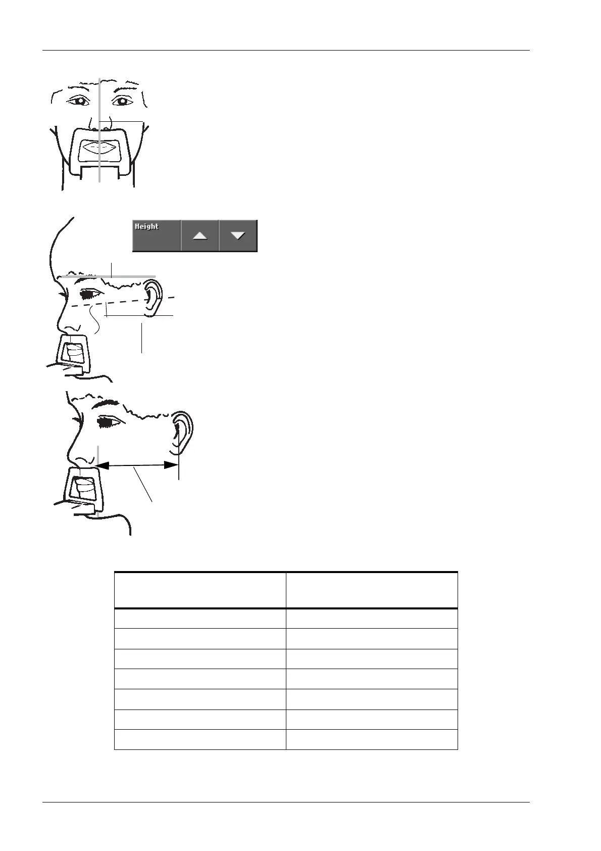
Position the patient’s head so that the midsagittal
plane coincides with the midsagittal plane light beam.
Make sure that the patient is looking straight ahead as
the light may appear to be correctly positioned but the
patient’s head could be turned slightly to one side.
Position the patient’s head so that the Frankfort plane
is tilted down 5°. To do this support the back of the
patient’s head with your hand and, using the Frankfort
plane light as a reference line, adjust the position of
the patient’s head by raising or lowering the vertical
carriage with the height adjusting keys. Make sure the
patient’s back is straight.
Measure the distance from the focal trough light beam
to the center of the auditory meatus and then
determine, from the table below, how much the patient
must be moved, backwards or forwards, to be correctly
positioned for the temporomandibular joint exposure.
POSITIONING GUIDE FOR TEMPOROMANDIBULAR JOINT EXPOSURES
Distance from light beam to
auditory meatus
Adjustment to the position
of the patient
80 mm +8 mm
85 mm +4 mm
90 mm 0 mm
95 mm -4 mm
100 mm -8 mm
105 mm -12 mm
110 mm -16 mm
Midsagittal plane light
Frankfort plane
Frankfort plane light
5°
Measure this distance