GE HEALTHCARE PROPRIETARY TO GE
D
IRECTION 5394227, 12 LOGIQ S8/LOGIQ E8 SERVICE MANUAL
Section 4-4 - Functional Checks 4 - 31
4-4-10 Tissue Velocity Imaging (TVI) Checks
4-4-10-1 Introduction
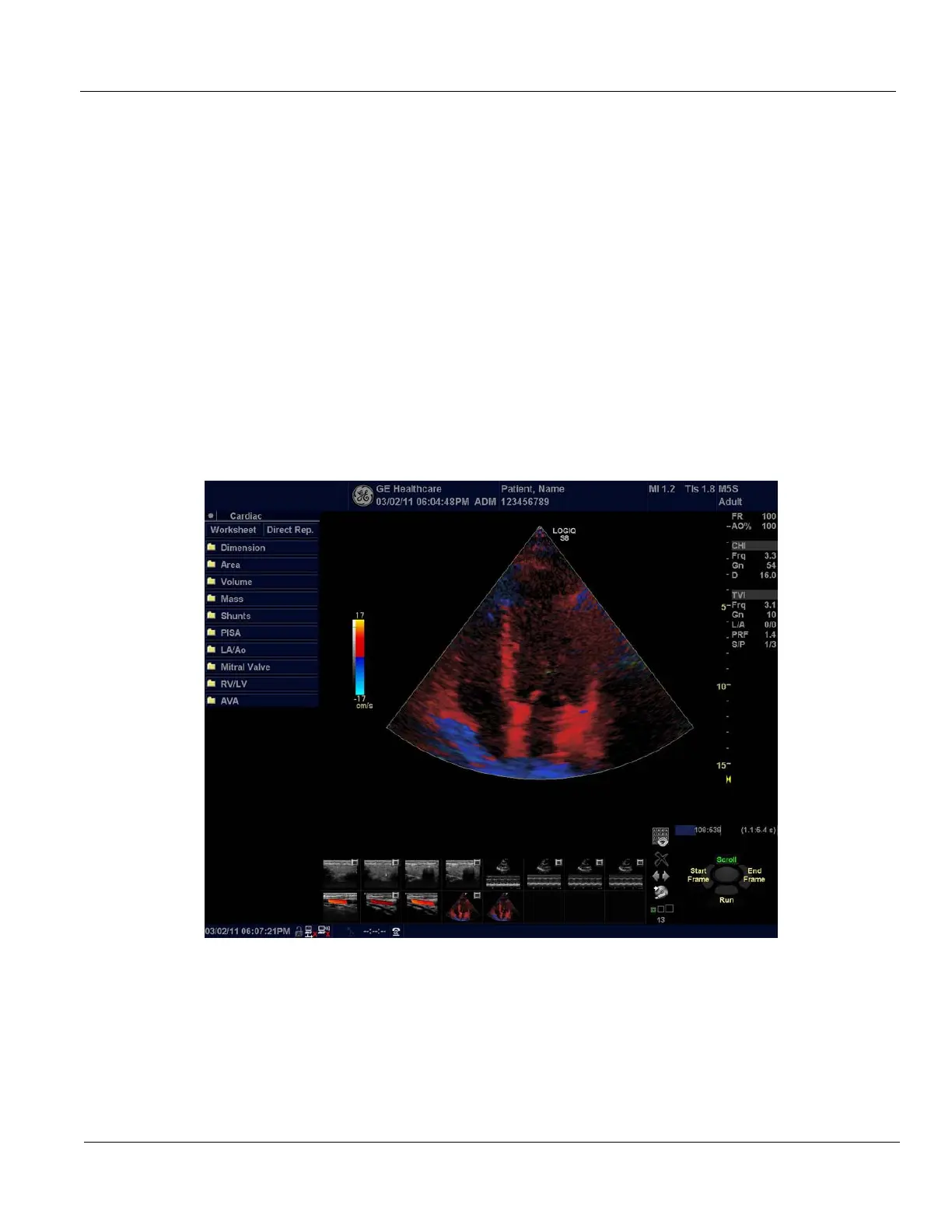
TVI calculates and color codes the velocities in tissue. The tissue velocity information is acquired by
sampling of tissue Doppler velocity values at discrete points. The information is stored in a combined
format with grey scale imaging during one or several cardiac cycles with high temporal resolution.
4-4-10-2 Preparations
1.) Connect one of the probes, to the scanner’s left-most probe connector.
-See: 3-6-4 "Connecting Probes" on page 3-15 for info about connecting the probes.
- For available probes, see: Section 9-13 "Probes" on page 9-59.
2.) Turn ON the scanner. The B-Mode window is displayed (default mode).
3.) If needed, adjust the Display’s Brightness and Contrast setting.
4.) Press TVI.
5.) To adjust the ROI size, press the top trackball key to select Size. To adjust the ROI position, press
the top trackball key to select Pos.
NOTE: If the trackball control pointer is selected, press trackball key to be able to select between
Position and Size controls.
Figure 4-25 TVI-mode Screen Example
 Loading...