24
JPK Instruments NanoWizard
®
Handbook Version 2.2
5. Cell imaging
The AFM has many advantages for cell imaging because
of the high resolution
and the ability to work in physiological conditions and even on living cells. Cells
are however at the extreme end of samples imaged with the AFM, since they are
so large and soft. Living cells are particularly challenging, since
the cell itself may
react to the imaging, and the stiffness is much less than for fixed cells. This
section gives some information to help get started with cell imaging, and recognize
the different effects that are seen when imaging living and fixed cells.
There are some important considerations when designing cell-
imaging
experiments, such as whether the cells are adherent or not, whether they should
be fixed or living, imaged in contact mode or intermittent contact mode and which
particular cantilevers
suit the experiment. Additionally, one must be aware of the
potential artifacts
that may arise during cell imaging, some of which are similar to
those observed in AFM images of other samples, some of which are unique to the
scanning of cells.
5.1 AFM in relation to other cell imaging techniques
Cells can be visualized by a number of different techniques, each generating
different information about cell structure and/or function. Obviously the
fundamental requirement of any imaging technique is that contrast
is somehow
generated. In terms of conventional optical microscopy this may be due to a
difference in material density (phase contrast), curvature (DIC) or with fluorescent
microscopy the emission of specific wavelengths of light from fluorophores
compartmentalized in specific locations
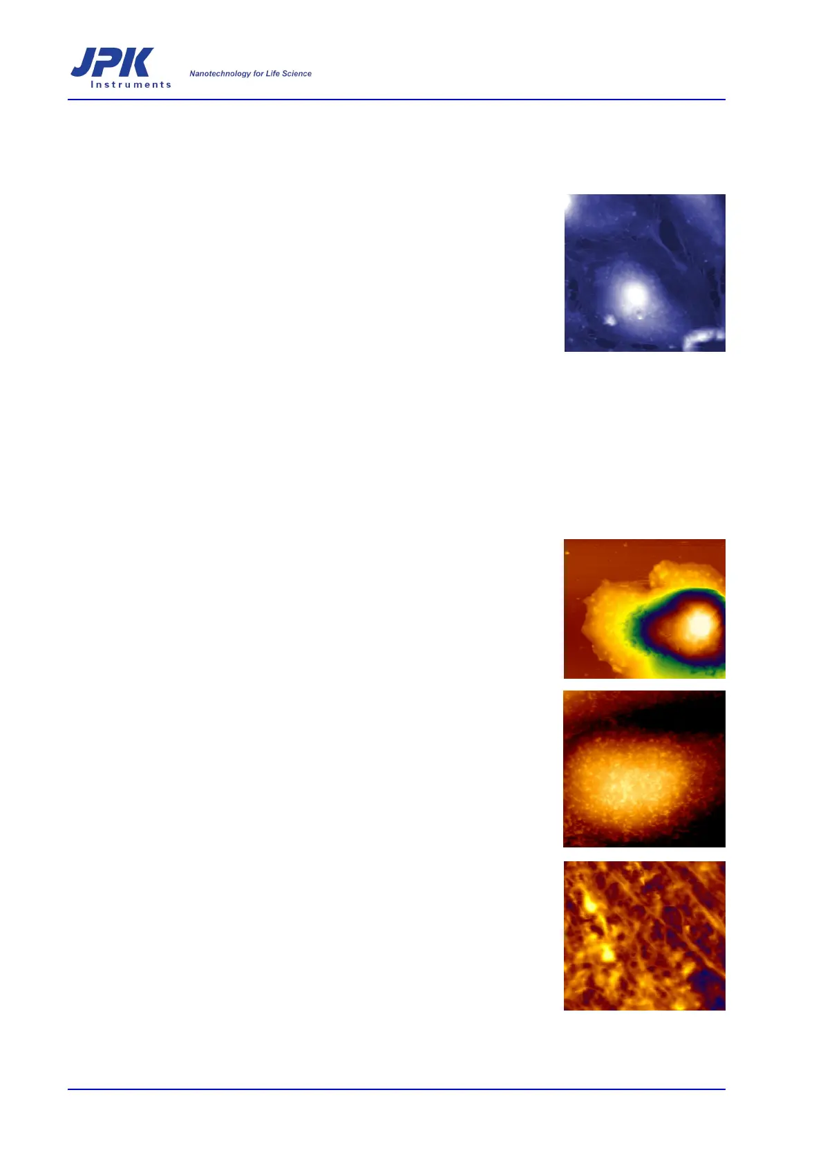
Contrast in atomic force microscopy imaging can be generated by a number of
sample properties. Topographic images from measuring the z-
based on height differences within a sample. An error signal image w
ill highlight
edges within the sample and in intermittent contact mode the phase image can
provide contrast based on material properties. Consequently, AFM imaging of cells
generates structural information. This leads to a number of possibilities in
experimental design. Large-
scale cellular movements can be monitored by
imaging living cells over time. Surface structure may be identified or the effect of
certain treatments on specific structure can be investigated. A major consideration
when imaging cells wi
th the AFM is the identification of functional components
within such structures, given the heterogeneity of the cell surface in terms of
protein composition and distribution. Consequently, while AFM imaging of cells can
generate novel information, the com
bination of AFM with other light microscopy
techniques expands the scope of possible experiments from structural studies, to
structure/function studies.