JPK Instruments NanoWizard
®
Handbook Version 2.2a
1
1. Introduction
1.1 About this handbook
Here you can find information about the principles and methods of scanning probe
microscopy. The focus is on applications in biotechnology and life science.
The particular details of the JPK NanoWizard
®
AFM system, for both the software
and hardware, can be found separately in the NanoWizard
®
User Manual. The aim
of this document is to introduce AFM for those who are not familiar with the
technique, and to provide background information and resources to
are familiar with the technique to extend their knowledge of particular applications.
The first sections of this handbook introduce the AFM technique, starting w
general ideas behind scanning probe microscopy. Later sections of this handbook
provide more detailed information and background for more experienced users.
1.2 What is an Atomic Force Microscope?
AFM is very different from optical microscopy.
Ther
e are no lenses, there is no requirement for a light source to illuminate the
sample, there is no eyepiece to look through; the microscope itself does not even
look like a typical optical microscope. The imaging technique consists of a
mechanical device, w
hich is able to measure very small forces when atoms or
molecules come close together, so it was named atomic force microscopy.
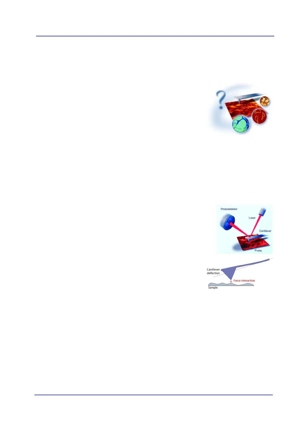
Cantilevers are at the heart of an atomic force microscope.
A critical part of the device called the cantilever is a plate sp
one end. At the other end it supports a pointed tip.
The tip can be moved across a
sample surface line by line, just like a lawn mower in the garden.
The pointed tip is brought into contact with the sample and moved across the
su
rface. The instrument measures the deflection of the cantilever as it scans, and
from this information about the tip movement a three-
dimensional image of the
sample is built up.