30
JPK Instruments NanoWizard
®
Handbook Version 2.2
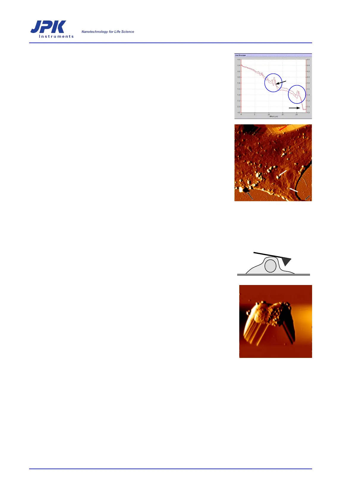
It can happen that the force applied to the cell is such that the tip starts to drift
away from the surface at
the edges (see arrows in image on the right) while still
displacing a structures (blue circles). It may be that the structures in question are
extremely flexible and will always be somewhat displaced. However, the user
could try either a) increasing the g
ains, or if this leads to oscillations b) decreasing
the scan speed and then decreasing the force.
In the image to the right some examples of scan faults can be seen. The image
presented here is an error signal image, 25 µm x 25 µm, taken in contact
mode in
the trace direction. The black arrows indicate areas of the scan where the tip was
not tracking over the surface due to too low an applied force. The white arrows
indicate some of the areas where protrusions have been displaced in the scan
direction. At the top of the image a red arrow indicates an unavoidable tip artifact
where the edge of the tip has been imaged as it slides down a steep surface.
5.6 Artifacts
General artifacts arising from normal tip geometry, damaged
contamination are described in Chapter 7. However, there is an additional artifact
that should be mentioned in the context of cell imaging. Imaging of cells can be
complicated by the height of the cell
body, particularly if the cell body is
significantly higher than other regions of the cell or substrate. If a comparatively
high structure is located under the legs of the cantilever, then the cantilever can
interact with the high features before the tip reaches the lower parts of the surface.
This artifact
can also occur if there are many dead, rounded cells in the sample. To
avoid interference from such structures the user should ensure that movement of
the cantilever does not bring the legs into contact
with any dead and rounded cells.
If the cells to be investigated have large height differences and an image cannot
be obtained with out this artifact
present then special cantilevers with a long tip
should be used, such as EBD tips (see Section 4.4).