JPK Instruments NanoWizard
®
Handbook Version 2.2a
3
Scanning Tunneling Microscope – STM
H. Rohrer, G. Binnig (1981)
The family of Scanning Probe Microscopes -
SPMs
Scanning Near-field
Optical Microscope
– SNOM
Photon Scanning
Tunnelling Microscope
- PSTM
Atomic Force Microscope
–
AFM
G. Binning, C. Quate, C
Gerber (1986)
Magnetic Force Microscope
- MFM
Electrostatic Force Microscope
- EFM
Shear Force Microscope
- ShFM
Scanning Ion Conductance
Microscope
- SICM
Scanning Capacitance
Microscope
- SCM
Scanning Chemical Potential
Microscope
-
SCPM
Scanning Thermal Microscope
- SThM
1.4 Atomic Force Microscopy
The atomic force microscope (AFM) is one of the fam
ily of scanning probe
microscopes, and is widely used in biological applications. The AFM uses a
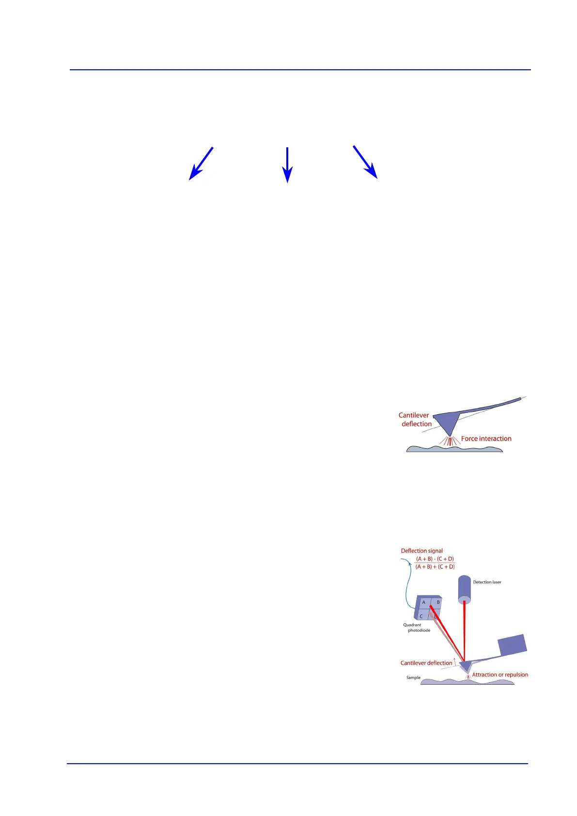
flexible cantilever as a type of spring to measure the force between the tip and the
sample. The basic idea of an AFM is that the local attractive or repulsiv
e force
between the tip and the sample is converted into a bending, or deflection, of the
cantilever. The cantilever is attached to some form of rigid substrate that can be
held fixed, and depending whether the interaction at the tip is attractive or
repulsive, the cantilever will deflect towards or away from the surface.
This cantilever deflection must be detected in some way and converted into an
electrical signal to produce the images. The detection system that has become
the standard method for A
FM uses a laser beam that is reflected from the back of
the cantilever onto a detector. The optical lever
principle is used, which means
that a small change in the bending angle of the cantilever is converted to a
measurably large deflection in the position of the reflected spot.
The attractive or repulsive force between the tip and the sample causes a
deflection of the cantilever towards or away from the sample. As the cantilever
deflects, the angle of the reflected laser beam changes, and the spot f
alls on a
different part of the photodetector. The signals from the four quadrants of the
detector are compared to calculate the deflection signal.
Most AFMs use a photodiode that is made of four quadrants, so that the laser spot
position can be calcula
ted in two directions. The vertical deflection (measuring the
interaction force) can be calculated by comparing the amount of signal from the
“top” and “bottom” halves of the detector. The lateral twisting of the cantilever can
also be calculated by comparing the “left” and “right” halves of the detector.
AFM is particularly suited for biological applications, because the samples can be