28
JPK Instruments NanoWizard
®
Handbook Version 2.2
5.4 Critical imaging parameters
Cantilever selection
Cantilevers with a spring constant between 0.01 N/m and 0.06 N/m can be used
for contact mode imaging of fixed cells. For contact mode imaging of living cells it
is better that the spring constant is as low as possible (0.01 –
softest cant
ilevers will be more susceptible to oscillations during scanning and may
require that lower gains are used.
Intermittent contact mode cantilevers can be somewhat stiffer, with a spring
constant of around 0.3 N/m. Triangular cantilevers are better than spr
ingboard
cantilevers for intermittent-contact mode imaging of cells.
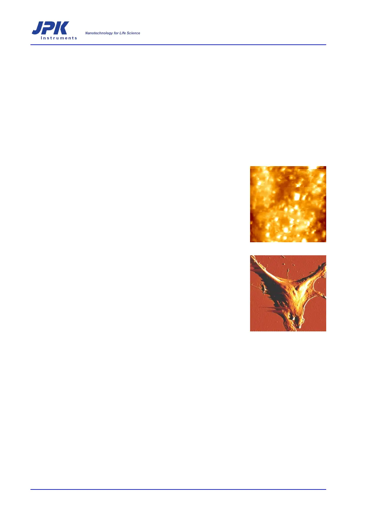
Setpoint selection
The selected set point should be just sufficient for the cantilever to stay in contact
with the surface. The set point should also be monitored over the course of the
scan to ensure that the applied force does not increase due to deflection drift. Too
high a force will lead to excessive displacement of surface structures in the scan
direction or tearing of the cell surface (see image to the right). Too low a force will
mean that the cantilever will not follow the contours of the cell sufficiently well.
(Note –
remember that increasing the setpoint in contact mode applies more force,
while in intermittent contact mode it corresponds to reducing the force, since it is
an amplitude)
Scan rate
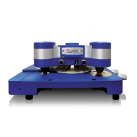
The line scan rate will mostly depend on the structure of the cells to be imaged.
Imaging of flat, well spread cells such as fibroblasts or endothelial cells will mean
that scan speeds of up to 5 Hz can be used. However, with cells
that have a high
cell body or other high features that have steep gradients at the edges, a
significantly slower scan speed may be required (even as slow as 0.2-
some living cells or cells that do not adhere well). If the scan speed is not reduc
in these cases the tip will fail to follow the surface correctly –
see image to the
right. The tip can be forced to follow the surface by increasing the applied force,
however, this will disrupt other structures at the surface of the cell – using a slow
er
scan rate achieves the same result without having a detrimental effect on other
features within the scan.
Error signal image of a fixed cell
imaged at too high scan rate in
contact mode
Drive amplitude in intermittent contact mode
When working in int
ermittent contact mode it is also important to set the drive
amplitude to an appropriate value. Too high a drive amplitude may lead to damage
of the cell as the cantilever drives into the surface (this will be apparent as the cell
will be pushed in the sca
n direction), too low a drive amplitude and the cantilever
may stick on the surface rather than freely oscillating. Generally a larger amplitude
is required for cell imaging than for other samples, such as single molecules. A
free amplitude of around 50 –
100 nm is a good starting range, though this may
need to be optimized for different cell types, depending how sticky or soft they are.
5.5 Using the oscilloscope to optimize the imaging parameters