34
JPK Instruments NanoWizard
®
Handbook Version 2.2
Generally well-
prepared single molecule samples are much less sticky than cells for
instance. Try an amplitude in the order of 5-15nm. If non-
calibrated amplitudes are
used, a
general sensitivity value of say 80 nm/V would give a suggested starting
amplitude of 400 mV –
1.2 V. Note that the driving frequency and setpoint must be
adjusted at proximity to the surface because the resonance and damping properties
may have changed close to the surface!
After a successful approach, the amplitude setpoint should be increased until the
contact with the surface is lost. After that, the setpoint should be decreased until the tip
is just tracking the surface. The gains can then be incr
eased to the maximum values to
get a good image. The scan speeds can be rather higher than for cell imaging, since
generally small regions are scanned, but remember the limitations discussed above
from the low cantilever resonance frequency. Line rates of
a few Hz would be typical for
scan regions around 1 micron.
6.3 Imaging hints - contact mode in liquid
In some circumstances, the best results can be obtained by imaging proteins using
contact mode. The absolute highest resolution images of proteins in li
quid tend to
come from protein crystals, particularly membrane proteins that form patches of protein
crystals that can be adsorbed well to mica. The proteins are stably stuck down, since
the area of such a membrane patch is so much larger than a single mo
proteins are extremely well-
supported laterally, so much less likely to be swept aside by
the AFM cantilever. Good results can also be achieved with contact mode in other
circumstances when the molecules are very well adsorbed to the surface.
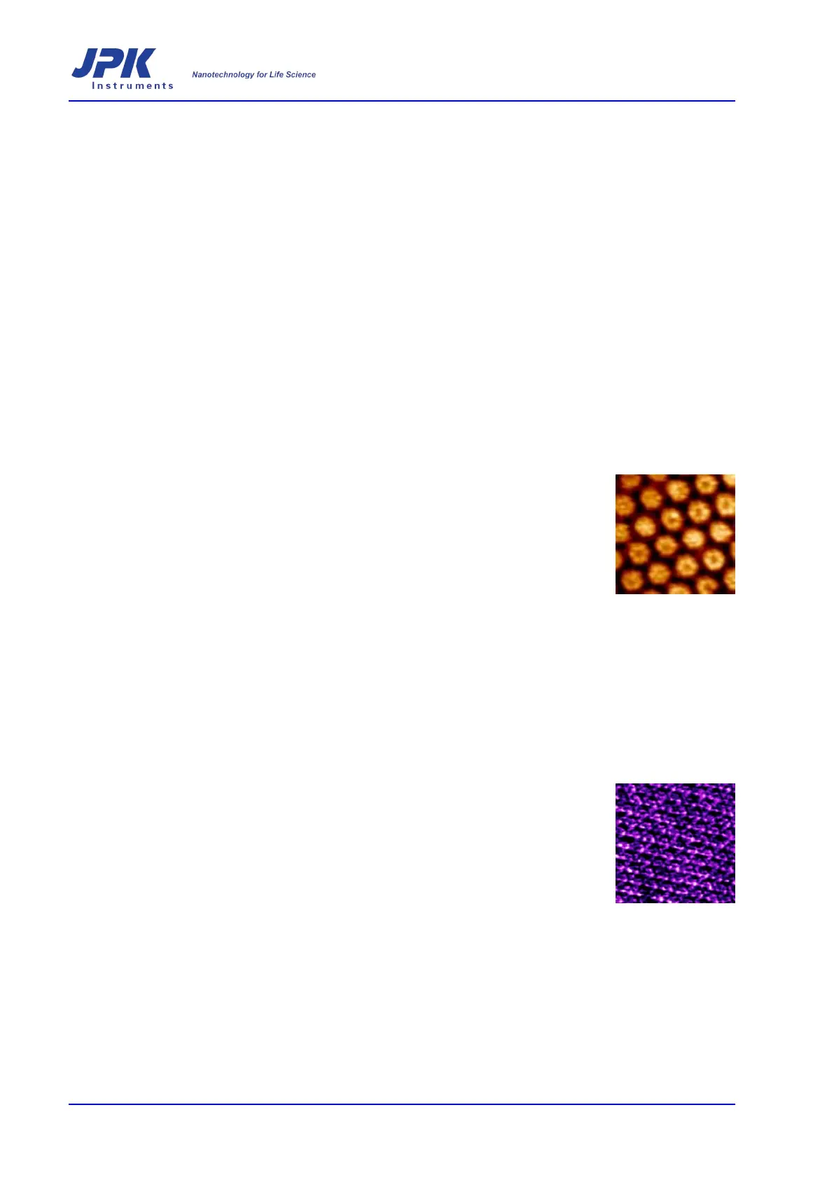
HPI membrane protein crystal
Cantilevers
Generally the softest possible cantilevers should be used, to reduce the lateral forces
applied to the molecules, for instance 0.03 N/m or less. In the case that the sample is
a lateral crystal, and the late
ral forces are not so critical, a slightly stiffer cantilever, for
example 0.06 N/m can have an advantage that the natural thermal noise fluctuations
are smaller, and this may improve the image slightly. As with intermittent contact
mode, a slightly smaller, more compact cantilever may also have advantages.
One consideration in contact mode is that any changes in the vertical deflection over
time directly affect the imaging force. To image with the lowest force, it is necessary to
minimize any changes
in the background deflection of the cantilever. Therefore using a
closed liquid cell may for instance help by reducing evaporation, which causes
temperature changes and ionic strength or pH changes that can cause the cantilever to
deflect. Uncoated silicon
cantilevers are much less sensitive to these changes than
silicon nitride, because a metal coating is necessary for silicon nitride cantilevers and
the different surface materials make them very sensitive to environmental changes.
Bacteriorhodopsin