26
JPK Instruments NanoWizard
®
Handbook Version 2.2
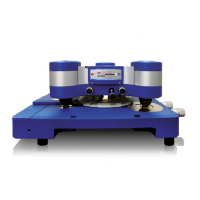
Typical images of living cells
Living fibroblast cells
The predominant feature of living
cells scanned in contact mode is the
subsurface cy
and height images shown)
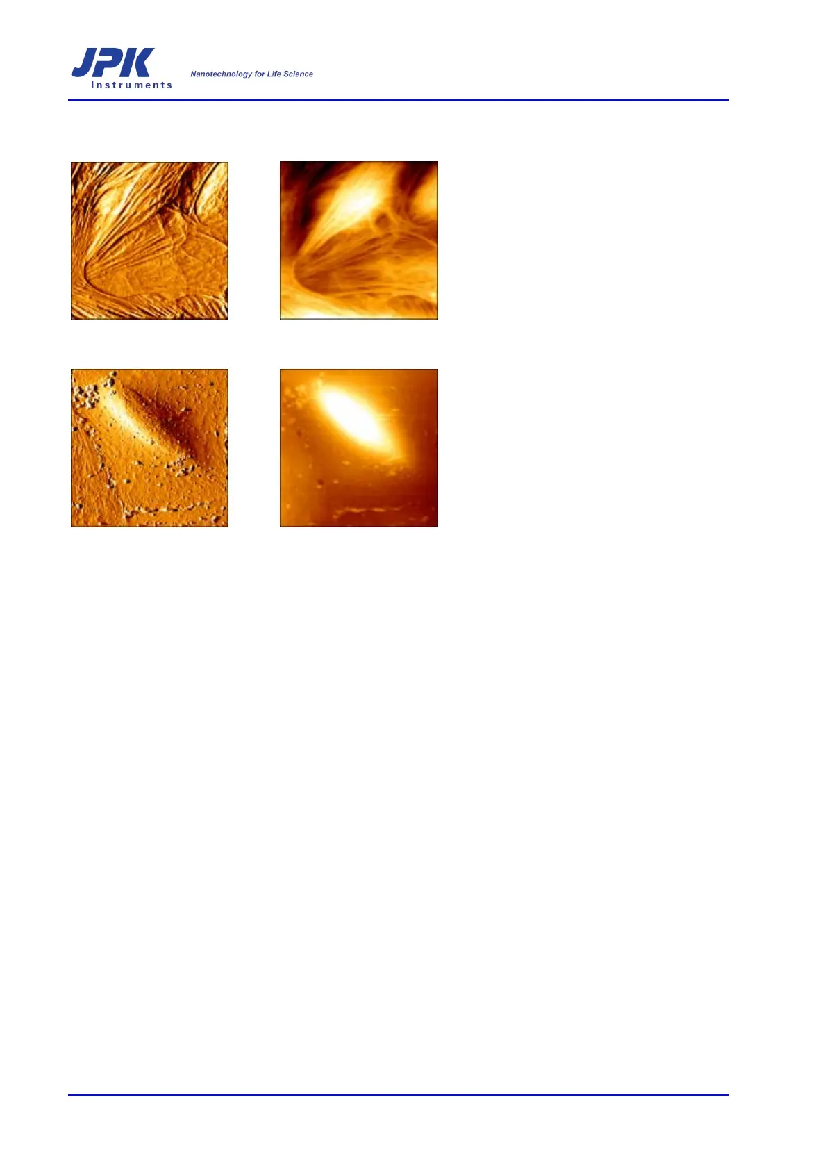
Typical images of fixed cells
Fixed fibroblast cells
Fixation of the cells leads to more
detailed information of smaller
surface (membrane) structures
cytoskeleton (deflection and height
images shown)
Choice of cantilever
The selection of the correct cantilever is critical for imaging cells. Selection of
which spring constant a cantilever should have will depend on which imaging
mode is to be used. Often very soft cantilevers are used fo
to minimize
the force. Sources of deflection drift, such as loss of liquid due to
evaporation, should also be minimized or the user will have to
adjustments to maintain a constant force. These considerations always exis
are more important when the force must be reduced as much as possible.
The use of unsharpened, as opposed to sharpened cantilevers is recommended –
particularly for imaging living cells. The potential achievable resolution is generally
reduced by u
sing an unsharpened cantilever, but for cell imaging the softness of
the cell surface will limit the resolution before tip size becomes an issue. The use
of unsharpened cantilevers reduces the chance of damaging the cell surface.
For simultaneous fluoresc
ence and AFM imaging it is recommended that a silicon
cantilever without a surface coating is used. The heat from the fluorescence lamp
leads to a deflection of the gold-
coated silicon nitride cantilevers, as these levers
are only coated on one side. If th
is happened during a contact mode scan there
would be a significant increase in force applied to the sample while the fluorescent
shutter was open. If the fluorescent images are taken between AFM scans, then
this is not such a problem, as the cantilever c
an be lifted off the surface while the
fluorescence shutter is open.