5-1
Chapter 5 ULTRASOUND (U/S) INSTRUCTIONS FOR USE
Overview
This chapter provides a step-by-step description for collecting tissue samples using the Mammotome revolve Dual
Vacuum-Assisted Biopsy System for an Ultrasound (U/S) Guided Procedure.
• SYMBOLS USED S
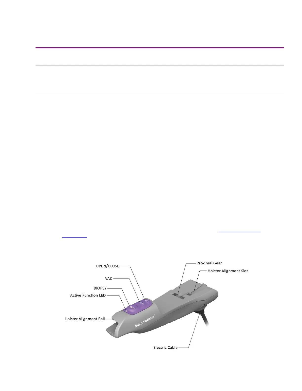
Mammotome revolve U/S Probe and U/S Holster
Summary Description
The Mammotome revolve U/S Probe and U/S Holster are used together to perform an ultrasound-guided breast
biopsy procedure.
• The holster is a non-sterile, reusable instrument with one electrical cable for transmitting information to the
control module. The holster communicates with the control module and U/S probe to drive the basic
functionality for the procedure. Buttons on the holster control the functions of the Mammotome revolve Dual
Vacuum-Assisted Biopsy System.
• The U/S Probe (with its integrated components) is a sterile, single-patient-use device that may be
used with imaging guidance to excise a tissue sample for diagnosis. The probe is designed to be
loaded onto the U/S holster. The probe consists of an outer trocar shaft and an inner cutter in a distal
sample aperture. The sample aperture can be manually rotated by using the wrist to rotate the
probe/holster assembly to the desired orientation. The probe comes packaged with an attached
vacuum tube set to connect the probe to a vacuum source. The tube set has three points of
attachment: 1) the control module vacuum connection slot, 2) the vacuum canister port, and 3) a
saline bag. For further convenience, the probe includes a Specimen Management System consisting
of 12 proximal specimen collection chambers. Each collection chamber is designed to capture and
hold excised tissue. The specimen collection chambers are designed to be removed from the fluid
management manifold in two separate trays (of six chambers each). If desired, the tissue can remain
in the chambers to be directly imaged in the post-biopsy specimen radiograph.
NOTE: U/S Probe and Holster Illustrations and Nomenclature are described in Chapter 2: System
Description.
Figure 21. U/S Holster Illustrated Nomenclature
 Loading...